Eye problems that are not defects of vision
In short-sightedness and long-sightedness, every part of the eye is healthy. The only trouble is that the eye brings light to a focus in front of the retina or behind it. A lens of the right kind in a pair of spectacles moves that focus back onto the retina, and the person sees clearly.
Some eye problems are of a different kind. In them, a part of the eye itself stops working as it should. The lens may turn cloudy, or special cells in the retina may fail. The light is not simply being focused in the wrong place, so spectacles alone cannot solve the problem. Three such problems are cataract, colour blindness and night blindness.
Cataract
The lens of the eye is clear, and light passes straight through it to the retina. The lens is made mostly of crystalline proteins. As a person grows older, these proteins begin to stick together, and the lens slowly turns cloudy, much like a glass window misted with steam. Light from objects can no longer reach the retina properly, so everything looks blurry.
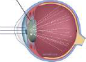
The cloudiness does not stay the same. As the lens grows more opaque, it lets less and less light through. In the end it loses its transparency completely, and the eye becomes fully blind. From the outside, the cloudy lens shows as a grey spot in the pupil, which in a healthy eye looks black.
Cataract: a grey-coloured spot that appears in the pupil of the eye because the lens has become cloudy.
A cataract is not a skin or film that grows over the front of the eye. It is the lens itself, sitting inside the eye behind the pupil, that turns cloudy. That is why the treatment is to take the lens out and replace it.
What raises the risk of cataract
Cataracts are mostly seen in elderly people, because growing old is the main cause. In some people, though, a cataract forms before old age. The chance of this is increased by:
- exposure to ultraviolet radiation from the sun
- cigarette smoking habits
- high diabetes, that is, too much sugar in the blood
Eye drops and spectacles cannot make a cloudy lens clear again. Cataracts are treated with surgery.
Cataract surgery
In cataract surgery, the cloudy lens is taken out of the eye and a clear artificial lens is put in its place. The work is done in this order:
- Step 1: A very fine hole is made at the edge of the cornea.
- Step 2: Ultrasound is sent in through the hole. Its vibrations break the cloudy lens into small pieces.
- Step 3: The pieces are taken out with the help of a vacuum tube.
- Step 4: An intraocular lens made of silicone or acrylic is placed where the old lens used to be.
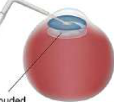

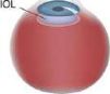
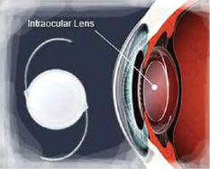
Intraocular lens (IOL): a small, clear artificial lens made of silicone or acrylic that is placed inside the eye in place of a cloudy natural lens.
The word intraocular simply means 'inside the eye'. The lens is a thin clear disc with two fine curved arms at its sides. The arms press gently against the inside of the eye and hold the lens steady. Because the new lens is clear, light once again passes cleanly to the retina after the operation, and the person can see.
Dr. Sanduk Ruit and the low-cost lens
For many years, intraocular lenses were costly, and many poor people with cataracts could not pay for the surgery. For them, a cataract simply meant going blind. The Nepali ophthalmologist Dr. Sanduk Ruit changed this. In 1995 he developed a very cheap intraocular lens, and this made cataract treatment cheap. People who could never have afforded the operation before could now have their sight given back.
Ophthalmologist: a doctor who specialises in the eye and its diseases.

How we see colour
The colour we see depends on two things: the light waves coming from an object, and the cells in the retina that identify colours. Sunlight is white light, a mixture of many colours. When it falls on a blue object, the object reflects the blue part of the light and absorbs the rest.
That reflected blue light enters the eye and is absorbed by the blue cone cells of the retina. The message that blue has been seen is then sent to the brain, and we see the object as blue. A red object works the same way, with red light and red cone cells.
The retina contains millions of cone cells of three types: blue cone cells, red cone cells and green cone cells. Between them, these three types separate one colour from another. The retina also holds a second kind of light-sensitive cell, the rod cell. Cone cells are short with a pointed tip, while rod cells are long and thin like a rod.
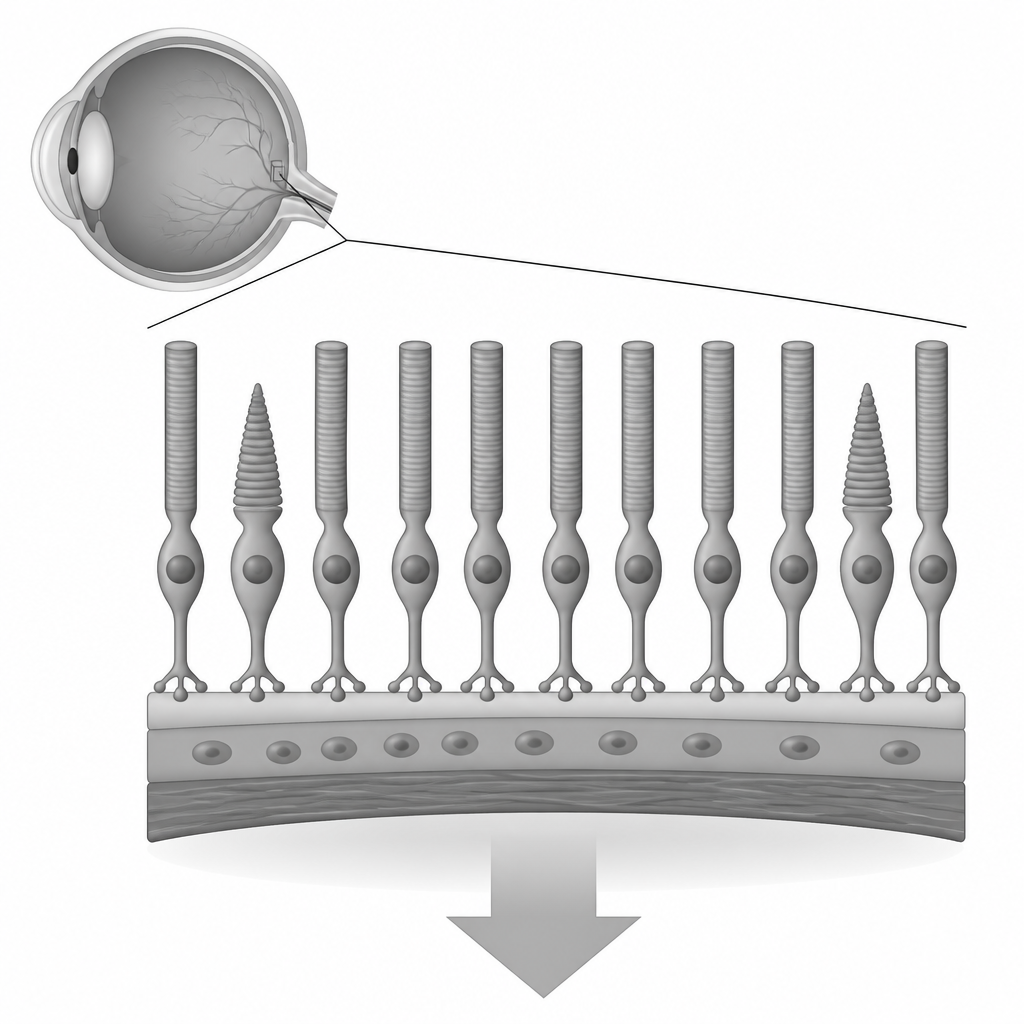
Cone cells: the cells of the retina that identify colours. There are three types: blue, red and green cone cells.
Colour blindness
If the cone cells in the retina stop working, or one type of them is faulty, a person cannot distinguish some colours. The commonest form is red-green colour blindness. A person with it cannot tell red and green apart, so a ripe red tomato and an unripe green one can look almost the same.
Colour blindness: the inability of the eyes to distinguish colours due to defects in the cone cells of the retina.
The main cause of colour blindness is heredity, which means it is passed down from parents to their children. Colour blindness can also occur when cone cells are destroyed by mutations or by harmful rays.
Colour vision is often checked with circles packed with coloured dots. The dots that form a number are one colour, and the dots around them are another colour of similar brightness. A person with normal colour vision reads the number at once. A person with red-green colour blindness may see only a jumble of dots, or read a different number.
Colour blindness almost never means seeing the world only in black and white. Most colour-blind people do see colours. They mix up certain ones, most often red and green.
Night blindness
Rod cells in the retina let us see in dim light. They are very sensitive to faint light, though they cannot tell colours apart. Thanks to them, if you walk into a room where the light is very dim, you can still make out where the bed, the table and the chairs are.
Sometimes the rod cells do not work properly. This can be due to disease, injury, lack of proper nutrients or many other reasons. The person then cannot see objects in very dim places. Unlike a cataract, this is a problem of the retina, not of the lens.
Night blindness (nyctalopia): the inability to see well at night or in dim places.
Rod cells contain a pigment called rhodopsin, which is made from a type of protein and vitamin A. When the body lacks vitamin A, it cannot make enough rhodopsin, and the rods stop responding to dim light. This is why a lack of vitamin A in the body is one of the main causes of night blindness. Night blindness can also be caused by heredity.
Rhodopsin: the pigment in the rod cells of the retina, made from a type of protein and vitamin A.
Vitamin A is found in foods such as carrots, green leafy vegetables, yellow fruits like mango and papaya, milk, eggs and fish.
Cones for colour, rods for dim light. Faulty cone cells cause colour blindness; faulty rod cells cause night blindness.
The three problems side by side
These three problems are easy to mix up, so it helps to set them side by side. Each one belongs to a different part of the eye, and each has its own main cause.
| Problem | Part of the eye affected | Main cause | What the person notices |
| Cataract | Lens | Old age; also ultraviolet rays, smoking and high diabetes | Blurred sight, a grey spot in the pupil, finally blindness |
| Colour blindness | Cone cells of the retina | Heredity; also mutations and harmful rays | Cannot tell some colours apart, most often red and green |
| Night blindness | Rod cells of the retina | Lack of vitamin A; also heredity | Cannot see well at night or in dim places |
The full lesson is waiting for you
The notes are free to read in full. A free account opens the rest of the practice, and tracks your progress and confidence score for every subject. Presentations and videos need approval from Learn Yeti.