The Tiny Units That Build a Body
Living things on the Earth run from creatures far too small for the naked eye to large, well developed animals and plants. An animal's body carries parts such as hands, legs, eyes, a stomach and a mouth. A plant carries roots, a stem, leaves, branches, flowers and fruits. Some less developed organisms have no such parts that a person can point at and name one by one. They are living things all the same, because they take food, they grow and they produce young ones of their own kind.

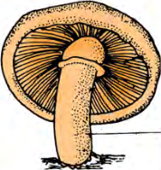

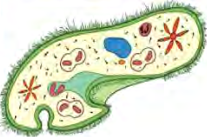
Every one of these parts and organs is itself built out of extremely small living cells. Inside one body there are lakhs upon lakhs of them, each doing its own share of work, and their work together is what keeps the different life processes running. That coordination is the reason a living body stays alive at all. Living beings are sorted into groups on the basis of how their bodies are built.
What a Cell Is
From a single celled microbe to a large many celled plant or animal, a cell is found in every living thing. Small or large, plant or animal, no living body is built without cells. A cell is the main base on which a body is built and on which every life process runs.
Cell: the smallest unit that builds the body of a living being and that carries out its life processes.
Digestion, breathing, reproduction, transport and photosynthesis all take place inside the cell, through chemical changes called biochemical reactions. Food looks as though it is digested by the stomach, but the work is actually done by the cells of the stomach. A leaf appears to carry out photosynthesis, but it is the cells inside the leaf that run the reaction.
The Cell as the Structural and Functional Unit
Because the life processes are carried out inside it, the cell is the functional unit of life. Because the body of a living being is built out of cells, the cell is also the structural unit of life. For these two reasons together, the cell is called the structural and functional unit of living beings.
A brick builds a house, so a brick is the structural unit of a house. But a brick does no work inside the house. A cell both builds the body and does the body's work. That is exactly why a cell is not only a structural unit but a functional one as well.
Cytology: the branch of science that studies cells.
The Discovery of the Cell and Cell Theory
Robert Hooke discovered the cell in the year 1665. He put a thin slice of cork under his microscope and saw that it was made of tiny rooms fitted together. He named those room like units with the Latin word Cellulae, which means a small room, and the name cell comes from that word. Hooke was born in 1635 and died in 1703.
Later, studies made by scientists such as Theodor Schwann, Matthias Jakob Schleiden and Rudolf Virchow showed that the bodies of all living beings are built of cells, and the idea that the cell is the basis of life came to be accepted.
Cell theory: the accepted idea that the bodies of all living beings are made of cells, and that the cell is the basis of life.
A Thin Section of a Leaf Stalk Under the Microscope
That cells are real is something a student can find out by looking, not only by reading. Cut a very thin section of the stalk of a green leaf, put it under a microscope, and small rooms fitted side by side come into view. The work is done in the order given below.
- Take a leaf of water hyacinth or of any other plant.
- Take the leaf stalk, which is called the petiole, and cut thin sections of it with a blade.
- Out of the many sections you have cut, choose the thinnest one.
- Place the chosen section on a clean glass slide.
- Cover it gently with a cover slip.
- Observe the temporary slide you have made under a microscope.
Cut sections with a blade only under your teacher's supervision. Always move the blade away from yourself and keep your fingers well behind the cutting edge.

Those small rooms seen in the section are the living cells. A single leaf stalk holds thousands of them fitted together, and it is these cells that give the stalk its shape and that carry water and food through it.
The Shape and Size of Cells
Cells come in many shapes. Animal cells are generally irregular in shape. They may be oval, round, rod shaped, long or cuboidal. A nerve cell is of the long kind, while a muscle cell is spindle shaped. Plant cells are mostly hexagonal, that is, six sided. Bacterial cells are round, rod shaped, long like a thread, or coiled like a spring, which is called a spiral shape.

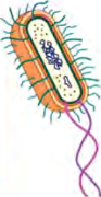
| Cell | Shape |
| Nerve cell | Long and drawn out |
| Muscle cell | Spindle shaped |
| Plant cell | Mostly hexagonal |
| Animal cell | Irregular: oval, round, rod, long, cuboidal |
| Bacterial cell | Round, rod, long thread like, spiral |
Do not think that all the cells of one organism have the same shape. A single human body holds long nerve cells, spindle shaped muscle cells and round cells at the same time, and each of them does a different job.
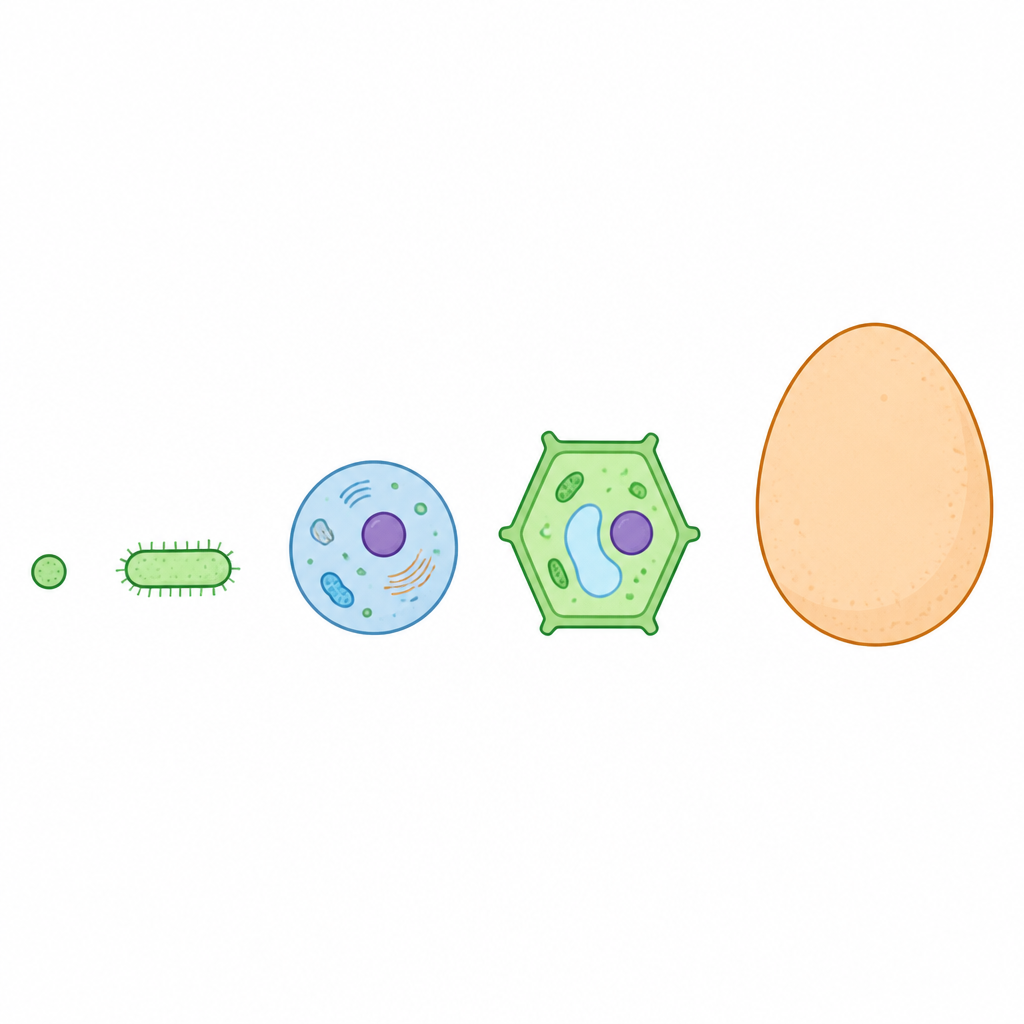
Cells range in size from ones far too small for the eye to ones that are quite large. The eggs laid by animals are cells too. The smallest cell is that of a bacterium named mycoplasma. It measures less than 1 micrometre, about 0.2 micrometre. The largest cell is the ostrich egg, which measures about 15 to 18 centimetres.
| Cell | Size |
| Smallest cell: the bacterium mycoplasma | About 0.2 micrometre |
| Most cells | Too small for the naked eye, a microscope is needed |
| Largest cell: the ostrich egg | 15 to 18 centimetres |
How a Cell Is Built
A cell is made of several different kinds of material. Its structure is generally divided into four parts. These are the covering, the cytoplasm, the cell components and the nucleus.
- Covering: the outermost boundary of the cell.
- Cytoplasm: the jelly like material that fills the space inside the covering.
- Cell components: the small bodies that sit inside the cytoplasm, each with its own job.
- Nucleus: the chief part that controls the work of the cell.
Making a Temporary Slide of Onion Cells
The purpose of this practical is to prepare a temporary slide of plant cells, observe it, and draw a labelled diagram of what is seen. The materials listed below are needed.
- An onion, or a piece of a leaf from any plant
- A blade and a needle
- A watch glass
- A slide and a cover slip
- Forceps and a brush
- Safranin and glycerine
- A microscope
Follow the method in the order given below.
- With forceps, peel off the thin skin of the onion or of the plant leaf, and cut it into small pieces with a blade.
- Using a brush, put those small pieces into a watch glass holding safranin.
- After a short while, move the pieces into another watch glass of water and wash them clean.
- Take a clean slide and put one drop of glycerine on it.
- With the needle, lay the thinnest piece of the peel into the glycerine and cover it with a cover slip so that no air bubble is trapped.
- The temporary slide of plant cells is now ready.
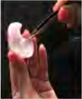
Never drop the cover slip straight down. Rest one edge on the needle and let it fall slowly from one side, or air bubbles will get in. Under the microscope such bubbles look like round shapes with a thick dark rim, and a student can easily mistake them for cells.
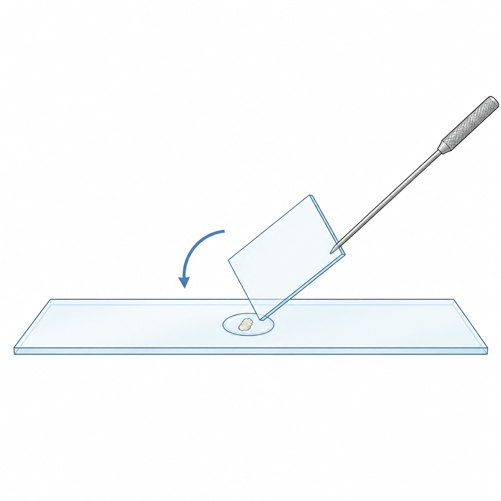
Place the finished slide under the microscope and observe it. Draw what you see in your practical book, marking the shape, the size, the parts and the special features of the cells, and show it to your teacher. Safranin stains the cells red, which makes the boundary of each cell and the darker body at its centre easy to pick out.
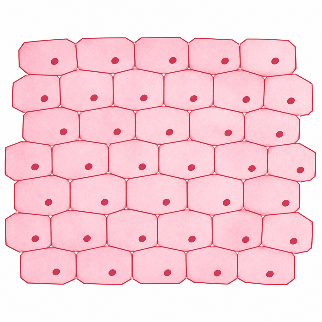
Instead of onion peel, this practical can also be done with the skin of a tomato fruit, the covering skin of a zebrina leaf, or the outer skin of an aloe vera leaf. Whichever peel is used, the thinner it is, the more clearly the separate cells show up.
Cells From the Inside of the Human Cheek
The purpose of this practical is to study the cells from inside a person's cheek. It needs a slide, a toothpick or a clean stick, a cover slip, a needle, methylene blue and a solution of sodium chloride. Follow the method in the order given below.
- With a sterilised toothpick or clean stick, gently and carefully scrape the inner surface of the cheek, so that the thin lining with its cells comes away.
- Put one drop of methylene blue and one drop of sodium chloride solution, which is common salt solution, at the middle of a slide, and place the thin scraping in it.
- Using the needle, cover the scraping with a cover slip so that no air is trapped.
Never use anything sharp or a metal needle to take the lining from the cheek. This practical must be done only under the supervision of a teacher.
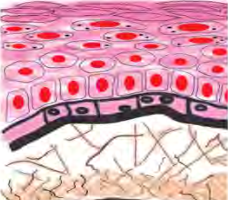
Place the slide under the microscope, observe it, and compare the shape, the size and the parts of these cells with the ones seen on the plant cell slide. Make a clean drawing of the cells and show it to your teacher. Instead of cheek lining, the thin inner layer of a hen's skin can also be used for this practical.
Comparing the Two Slides
Looking at the two slides one after the other makes something plain without being told: cells are found everywhere, yet they are not all alike. The comparison below shows what to look for while observing.
| What to look at | Onion peel cells | Cheek cells | |
| Shape | Regular, fitted like bricks | Irregular and flat | |
| Outer boundary | Clear and straight edged | Soft and wavy | |
| Arrangement | Packed in rows, touching each other | Scattered and separate | t |
| Stain used | Safranin | Methylene blue |
Under the microscope the shapes differ, but inside each cell there is one darker round body. That is the nucleus, and it is found in the cells of plants and of animals alike.
The full lesson is waiting for you
The notes are free to read in full. A free account opens the rest of the practice, and tracks your progress and confidence score for every subject. Presentations and videos need approval from Learn Yeti.