Living Things the Eye Cannot See
Take an egg, crack the shell a little on one side, stand it in a beaker and leave it alone for a few days. When you come back four or five days later, the material inside has gone bad and an unpleasant smell is rising from it. Nobody touched the egg and nobody added anything to it, yet it spoiled by itself. The change was brought about by extremely small living things that arrived from the air and from the surroundings. You cannot see them with your eyes, but you can see their work in the rotting egg and smell it in the air.
Never taste or eat the rotted egg, and do not put your nose close to it to smell it. When the observation is over, throw it into the rubbish and wash your hands well with soap and water.
Many kinds of living things share the world with us. Some of them are large, like an elephant or a pine tree, and you can pick them out from a distance. Others are so small that no amount of staring will show them to you. To look at these you need a microscope.
A living thing so small that it cannot be seen with the naked eye is called a microorganism.
Microorganisms affect our daily life all the time. The egg going bad and giving off a smell was their work. There is a whole branch of science that studies nothing but these creatures.
The branch of science that studies microorganisms is called microbiology.
Where They Live and Which Ones Are Microorganisms
Microorganisms are not tied to one kind of place. They are found in air, in water and in soil. They live on the food we eat, on the pots we use and on our own skin. Even in places far too hot or far too cold for a large animal to survive, some microorganism or other is found. That is why they count as the most widely spread group of living things on earth.
The living things listed below are all examples of microorganisms.
- Bacteria
- Some fungi, such as yeast and slime mould
- Some algae, such as Chlamydomonas and Chlorella
- Protozoans, such as amoeba, paramecium, plasmodium and vorticella
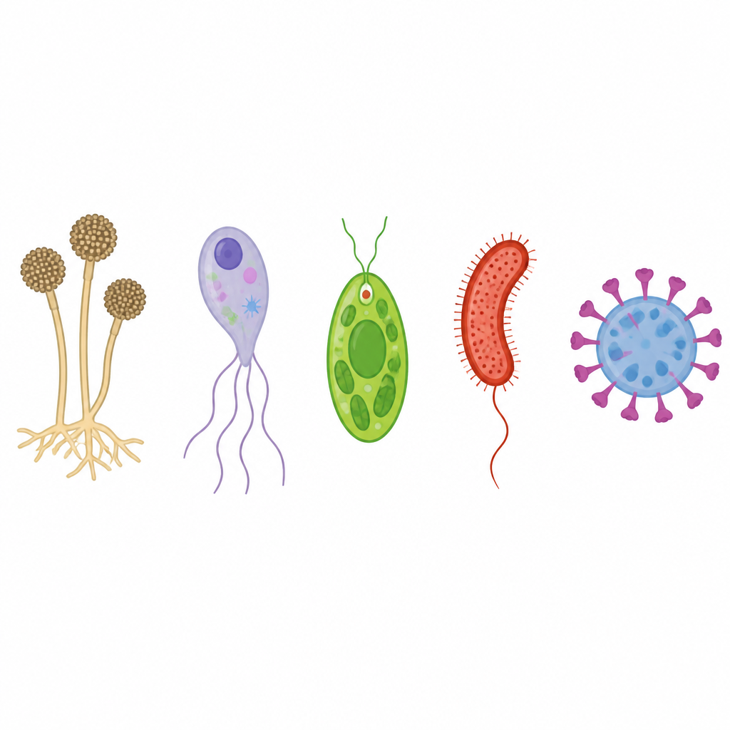
Microorganisms do not all come from one kingdom. Bacteria belong to Monera, amoeba belongs to Protista and moulds belong to Fungi. Being small is a matter of size, not a group in the classification system.
Four of these groups are worth looking at closely. Their build, their movement and the way they feed are quite different from one another.
Virus: a tiny particle with no cell at all. It multiplies only inside a host cell.
Bacteria: single-celled living things of the Monera kingdom. They come in round, rod and thread-like shapes.
Amoeba: a single-celled living thing of the Protista kingdom. Its shape is irregular and it moves on pseudopodia.
Fungi: living things built from a web of mycelium. They take their food from rotting matter.
(a) Virus
A virus is a tiny disease-causing particle. Its build is unlike that of every other living thing, because a virus has no cell. Inside it there is a single strand of DNA or RNA, and that strand is wrapped from outside in a coat of protein. Since it has no cell, a virus is not called a living organism but a tiny particle.
A virus is a tiny particle that has no cell and is made of a strand of DNA or RNA covered by a coat of protein.
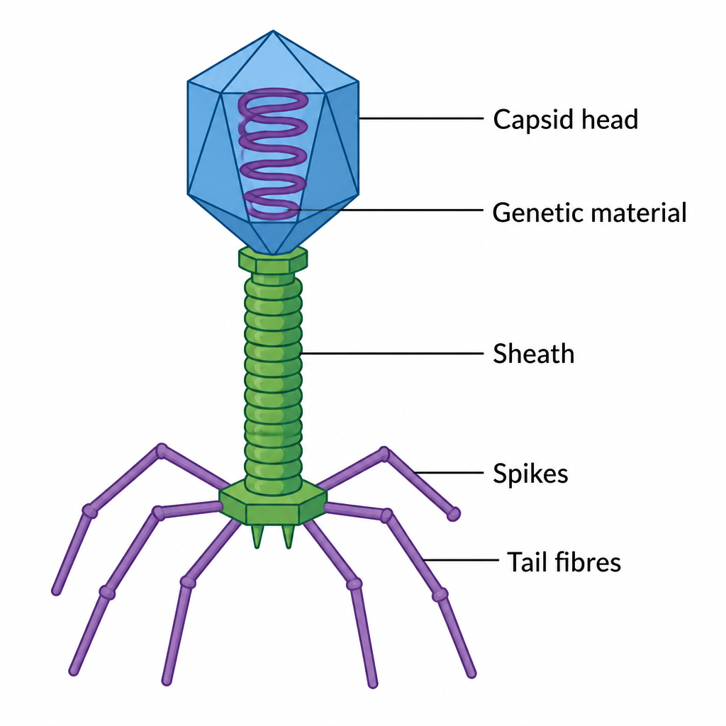
A virus shows the properties of both living and non-living things. Out in the open it behaves like a non-living object. It does not feed, it does not grow and it runs no life process at all. But once it enters a host cell it starts multiplying rapidly, just as a living thing would.
Because a virus must take every part it needs for running its life processes from the host organism, it is called an obligatory parasite.

Viruses are found everywhere, in air, in water and on land. Rhino virus, corona virus, tobacco mosaic virus and bacteriophage are examples of viruses.
Do not think of a virus as a small bacterium. A bacterium has a cell and a virus has none, so calling a virus a single-celled organism is wrong.
(b) Bacteria
Bacteria are microorganisms of the Monera kingdom. The body of a bacterium is made of one single cell, so bacteria are single-celled organisms. They are not all the same shape. Some are round, some are rod shaped and some are long and thread-like.
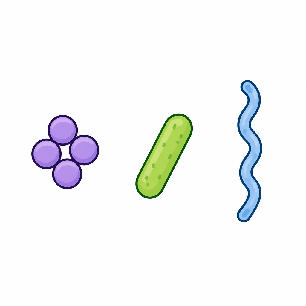
Bacteria are found in air, in water and in soil, and also in harsh conditions such as very hot or very cold places. Their cell is of the prokaryotic type, and it is covered by a strong covering called a capsule. This tough covering is one reason bacteria manage to protect themselves even in difficult surroundings.
The strong covering that wraps a bacterial cell from outside is called a capsule.
Bacteria do not all get their food in one way. Some make their own food by photosynthesis. Some make it by chemosynthesis. Others are parasites and take their nourishment from the body of another living thing. Cyanobacteria, Rhizobium, Salmonella, Azotobacter and Pseudomonas are examples of bacteria.
(c) Amoeba
An amoeba is a single-celled microorganism of the Protista kingdom. It is found in water, in soil and on rotting matter, and some kinds live inside the body of another animal as parasites. Its body has an irregular shape, so an amoeba has no fixed form.
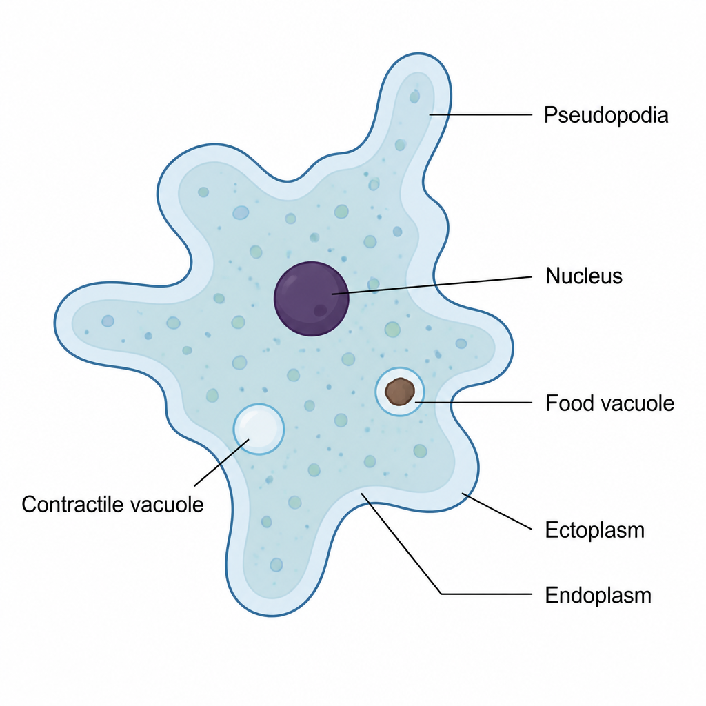
An amoeba moves with the help of pseudopodia. The fluid part of its body pushes out on one side and the rest of the body is drawn after it, and in this way the amoeba creeps along. What is worth noticing is that the amoeba runs all its life processes, nutrition, digestion, excretion and reproduction, with that one single cell. In a large animal separate organs do these jobs, but in an amoeba one cell carries the whole load.
The temporary foot-like part that an amoeba pushes out of its body to move and to catch food is called a pseudopodium, and more than one are called pseudopodia.
There are many kinds of amoeba and not all of them cause disease. Two kinds, however, are harmful to human beings.
- Entamoeba histolytica is found in the human intestine and causes the dysentery known as amoebiasis.
- Naegleria fowleri lives freely in water, but when it gets into the human body it too causes disease.
(d) Fungi
The living things of the mould group are fungi. They belong to the kingdom Fungi, also written as Mycota. Fungi usually grow on things that are rotting and take their food from there. That is why mould appears on damp bread, on spoiling fruit and on wet wood.
A living thing that takes its nourishment from dead and decaying matter is called a saprophyte. Most fungi are saprophytes.
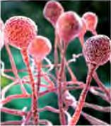
Not every fungus is a saprophyte. Some fungi are parasites and infect animals and plants, causing disease in them. The body of a fungus is built from mycelium, which is made of fine threads like strands of cotton. Even the cap and stalk of a mushroom are nothing but these threads packed tightly together.
The web of fine cotton-like threads that makes up the body of a fungus is called mycelium.
Mucor, mushrooms, yeast, yarsagumba which is the caterpillar fungus of the high Himalayan pastures, penicillium and puccinia are examples of fungi. Among these, yeast cannot be seen without a microscope while a mushroom can be spotted from across a field. So not every member of the fungi kingdom is a microorganism, only some of them are.


Never pick and eat a mushroom that has come up on its own in a field, a forest or beside a path. Many mushrooms are poisonous, and colour and shape alone cannot tell you which ones are safe.
The Four Groups Side by Side
Putting the main differences between the four groups in one place makes them easier to hold in mind.
| Group | Body structure | Key feature | Examples |
| Virus | No cell, a strand of DNA or RNA in a protein coat | An obligatory parasite that multiplies only inside a host cell | Corona virus, bacteriophage |
| Bacteria | One prokaryotic cell covered by a capsule | Round, rod and thread shapes, able to survive harsh conditions | Rhizobium, Salmonella |
| Amoeba | One cell of irregular shape | Moves on pseudopodia, runs every life process in one cell | Entamoeba histolytica |
| Fungi | Mycelium made of fine threads | Saprophytic, and some are parasitic | Mucor, mushroom, yeast |
Looking at Mucor Under a Microscope
Reading about mycelium is not the same as seeing it, and it is only after you have looked at it that the idea settles. For this you need a potato, a slide, cotton blue stain, a brush, a coverslip, a beaker, a watch glass and a microscope.
- Put a piece of bread or a little soaked beaten rice in a watch glass and leave it in the laboratory for a few days.
- After a few days, look closely at the mucor that has grown on it.
- Place one drop of cotton blue stain in the middle of the slide.
- With the help of the brush, gently lift the mycelium of the mucor and lay it in the cotton blue.
- Lower the coverslip slowly over it so that no air is trapped underneath.
- Put the finished temporary slide on the microscope, observe it, and draw the structure you can see.
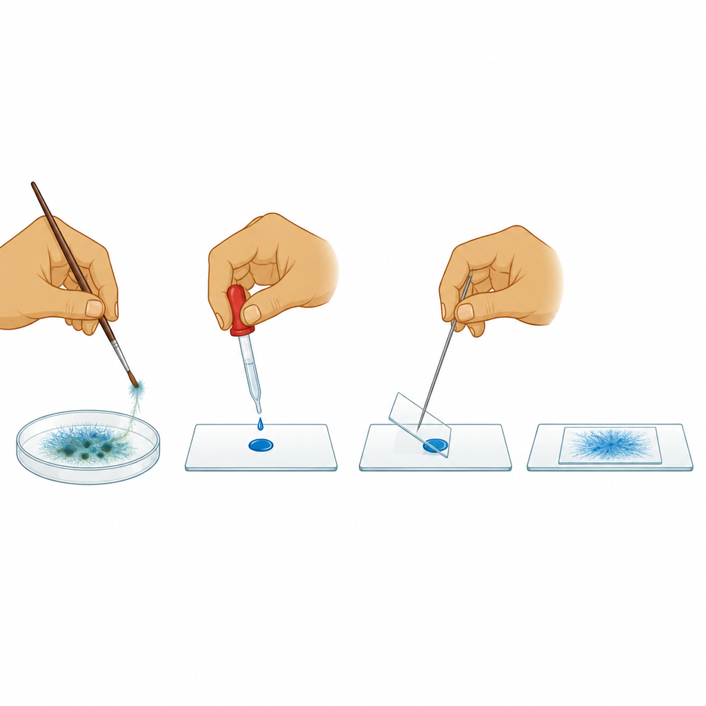
Under the microscope you see thread-like strands stained blue, and here and there small round heads at the tips of those strands. The web these threads make together is the mycelium. In the same way, permanent slides of amoeba, paramecium and yeast can be placed under the microscope and studied, and their features drawn.
Never taste the mouldy bread or beaten rice, and do not hold it close to your nose to smell it. Slides and coverslips are made of glass, so handle them carefully. Wash your hands with soap and water when the work is finished.
The full lesson is waiting for you
The notes are free to read in full. A free account opens the rest of the practice, and tracks your progress and confidence score for every subject. Presentations and videos need approval from Learn Yeti.