Body Systems and the Waste the Body Makes
The human body is built in layers. Tiny cells group together to form tissues. Tissues group together to form organs such as the heart, the lungs and the kidneys. Organs that share one job form a system, and all the systems together make up the whole body. The skeletal, muscular, digestive, respiratory, circulatory and nervous systems each have their own work, yet the body stays healthy only because they work in step with one another.
Cells → tissues → organs → systems → the human body
After food is digested, the body absorbs the nutrients it needs. When cells use those nutrients to do their work, they also produce substances the body has no use for, and some of these are harmful. These are called waste substances. At times the body also has more water or salt than it needs. All of this travels round the body in the blood. If it is not separated from the blood and thrown out, the body becomes ill. The urinary system does this large cleaning job.
What the Urinary System Is
Urinary system: the system that removes waste substances from the body in the form of urine, which is made by the kidneys.
No single organ does this job alone. Four organs work one after another, and urine passes from each to the next until it leaves the body. The organs of the urinary system are:
- Kidneys: filter the blood and make urine.
- Ureters: carry urine from the kidneys to the bladder.
- Urinary bladder: stores urine until it is passed out.
- Urethra: carries urine from the bladder to the outside of the body.
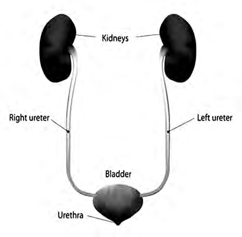
Follow the path of urine in the diagram. Urine is made in both kidneys at the top, runs down the two thin ureters, collects in the bladder and finally leaves through the urethra. There are two kidneys and two ureters, but only one bladder and one urethra.
The Kidney: Shape, Size and Position
The kidney is a dark red organ shaped like a bean seed. There are two kidneys. They lie inside the abdomen, below the diaphragm and close to the waist. One kidney is on the right side of the backbone and the other is on the left. They are not at exactly the same height: the left kidney sits a little higher than the right one.
| Feature | Detail |
| Shape | Like a bean seed |
| Colour | Dark red |
| Number | 2 |
| Length | 11 cm |
| Width | 6 cm |
| Thickness | 3 cm |
| Weight | 130 g to 150 g each |
| Position | In the abdomen, below the diaphragm, one on each side of the backbone |
The outer edge of each kidney bulges outward, which is called convex. The inner surface curves inward a little, which is called concave, and this dip faces the back, towards the backbone. The dip is called the hilum, and it works like the kidney's doorway. The renal artery enters the kidney through the hilum, bringing blood to be cleaned. The renal vein leaves through the hilum, taking the cleaned blood away, and the ureter leaves from here too.
Hilum: the dipped inner part of the kidney, through which the renal artery enters and the renal vein and the ureter leave.
An adrenal gland sits on top of each kidney like a cap. Although it rests on the kidney, it is not part of the urinary system and takes no part in making urine.
Inside the Kidney: Cortex, Medulla and Renal Pyramids
If a kidney is sliced from top to bottom so that its front half and back half separate, two regions can be seen clearly. The outer region looks reddish brown, and the inner region is paler. Inside the pale region there are several triangle-shaped bundles.
Cortex: the outer, reddish brown region of the kidney.
Medulla: the inner, paler region of the kidney.
Renal pyramids: the triangle-shaped bundles of fibres found in the medulla.
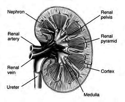
Beyond the tips of the pyramids, near the hilum, there is a wide hollow space called the renal pelvis. All the urine made in the kidney gathers here first and then drains into the ureter. The real work of making urine, though, is done by extremely tiny tubes spread through the cortex and medulla. These tubes are called nephrons.
The Nephron: the Working Unit of the Kidney
When a kidney is examined under a microscope, it shows hundreds of thousands of tiny tube-shaped structures. These are nephrons. Each nephron is like a small factory that cleans blood and makes urine. Because so many of them work at the same time, the kidneys can clean all the blood in the body.
Nephron: one of the hundreds of thousands of tiny tube-like structures in the kidney. The nephron is the functional unit of the kidney, its smallest working unit.
The nephron begins in the cortex. There, tiny blood vessels are coiled into a tangled ball, and the cup-shaped beginning of the nephron wraps around it. This tangled ball of blood vessels is called the glomerulus. These vessels bring blood to the nephron, and this is where the blood is filtered. The cup around the glomerulus is the glomerular capsule, labelled Bowman's capsule in the diagram.
Glomerulus: a tangled ball of tiny blood vessels in the cortex, which supplies blood to the nephron and where the blood is filtered.
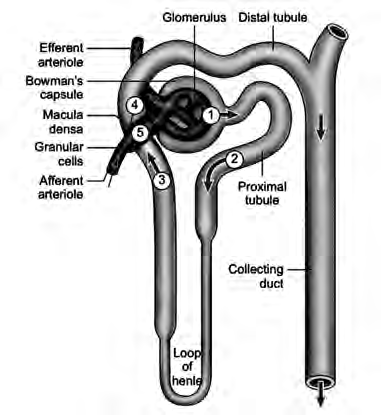
From the capsule, a long winding tube begins. It twists, dips down, comes back up and finally joins a collecting tubule. The liquid filtered out at the glomerulus is not all waste. It also holds water and other substances the body still needs. So, as this liquid flows along the winding tube, most of it is soaked back into the blood. This is called reabsorption. Only the small part that is left becomes urine and passes into the collecting tubule.
Reabsorption: the process in which the useful part of the liquid filtered at the glomerulus is taken back into the blood from the nephron's tube.
The collecting tubules of many nephrons join one another as they go and finally open into the renal pelvis. From there the urine drains into the ureter.
Numbers show how big this job is. Together, the two kidneys filter about 180 litres of liquid every day. Yet only about 1.5 litres of it leaves the body as urine. The remaining 178.5 litres or so is reabsorbed and mixes back into the blood. Without reabsorption, the water and useful substances the body needs would be lost along with the urine.
About 180 litres filtered each day, about 1.5 litres passed as urine. Everything else is reabsorbed into the blood.
Functions of the Kidney
Making urine is only one of the kidney's jobs. While it filters the blood and takes part of it back, the kidney is also keeping the inside of the body in balance. Its main functions are:
- It makes urine.
- It keeps the amount of water in the body balanced.
- It filters the blood and regulates blood pressure.
- It keeps the amount of salts in the body balanced.
- It keeps the amount of plasma in the blood steady, and so keeps blood pressure right.
- It throws out substances that are useless or in excess, through urine.
You can see the water balance in everyday life. On a hot day when you sweat a lot, the body loses water. The kidneys then take more water back into the blood, so you pass less urine and it looks darker. On a day when you drink plenty of water, the kidneys let the extra water go, so you pass more urine and it looks paler.
The Ureters
A thin tube leaves the hilum of each kidney and runs down to the bladder. This tube is the ureter. Since there are two kidneys, there are two ureters. A ureter is a muscular tube, 25 to 30 cm long and 3 mm thick. Its main job is to carry the urine made in the kidney down to the urinary bladder.
Ureter: the muscular tube that carries urine from the kidney to the urinary bladder.
Ureter and urethra sound alike but they are different tubes. There are two ureters, and they bring urine from the kidneys to the bladder. There is only one urethra, and it takes urine from the bladder out of the body.
The Urinary Bladder
The urinary bladder is a muscular sac. Its wall has three layers and is made of elastic muscle, so it stretches like a balloon as it fills with urine. It lies in the lower abdomen and is shaped like a pear. Urine brought by both ureters collects here, which makes the bladder the body's store of urine. The urethra is joined to its lower tip.
An adult's bladder can hold about 500 millilitres of urine, and it can stretch to hold up to about 700 millilitres. But we know long before it is full. Once about 200 to 300 millilitres has collected, we begin to feel the need to pass urine, and by about 300 millilitres the feeling is clear.
The Urethra
The urethra is a single tube that runs from the lower tip of the bladder to the outside of the body. It carries the urine stored in the bladder out of the body. It is different in males and females.
Urethra: the tube leading out of the bladder, which carries urine out of the body.
| Feature | Male | Female |
| Length | About 18 cm | About 4 cm |
| Where it opens | At the tip of the penis | Just above the opening of the vagina |
| What passes through it | Both urine and semen | Urine |
At both the upper and the lower end of the urethra there is a urethral sphincter, a ring of muscle. It keeps the urethra squeezed shut at all times except when we pass urine, so urine does not leak out on its own.
Urethral sphincter: the ring of muscle at the ends of the urethra that keeps it closed except when urine is being passed.
How Urine Leaves the Body: Micturition
Micturition: the process by which urine, made in the kidneys and stored in the bladder, is passed out of the body through the urethra.
The process has two parts. In the first, urine is made in the kidney and reaches the bladder. In the second, the bladder is emptied after a signal from the brain. The whole sequence runs like this:
- Blood is filtered at the glomerulus into the glomerular capsule, and the filtered liquid moves on into the renal tubule.
- In the renal tubule, much of the water, sodium, glucose, protein and similar substances is reabsorbed into the blood.
- What is left after reabsorption is urine. It flows through the tubules into the renal pelvis.
- As soon as urine is made, it runs down the ureters and collects in the bladder.
- When about 300 ml of urine has collected, the bladder swells, and sensory nerves in its wall tell the brain that it is filling.
- The person feels the urge to pass urine.
- When the brain signals the bladder and the urethra, the sphincter muscle where the two meet opens and the muscle of the bladder squeezes.
- The urine stored in the bladder flows out of the body through the urethra.
It is the brain that learns the bladder is full and gives the order to empty it. Sensory nerves carry the message in, the brain sends the signal out, then the sphincter opens and the bladder squeezes.
Looking After the Urinary System
The waste the kidneys take out of the blood leaves the body dissolved in the water of urine. So this system works well only when the body has enough water. We should drink about 3 to 5 litres of water a day.
Do not hold urine in for a long time. When you feel the need to pass urine, go to the toilet without delay.
The Four Organs at a Glance
| Organ | Number | Size | Main job |
| Kidney | 2 | 11 cm long, 6 cm wide, 3 cm thick | Filters blood and makes urine |
| Ureter | 2 | 25 to 30 cm long, 3 mm thick | Carries urine to the bladder |
| Urinary bladder | 1 | Holds about 500 ml | Stores urine |
| Urethra | 1 | About 18 cm in males, about 4 cm in females | Carries urine out of the body |
The full lesson is waiting for you
The notes are free to read in full. A free account opens the rest of the practice, and tracks your progress and confidence score for every subject. Presentations and videos need approval from Learn Yeti.