The Circulatory System: Heart, Blood and Blood Vessels
Every cell in your body needs a steady supply of oxygen and food to stay alive and do its work. The same cells also make carbon dioxide and other waste, which must be carried away quickly. Blood does this carrying. Blood cannot move by itself, though. It needs a pump to push it and a set of tubes to flow through. The heart is the pump, and the blood vessels are the tubes.
Circulatory system: the system in our body that keeps the blood flowing. It is made up of the heart, the blood and the blood vessels.
The first person to study this system was William Harvey. Its main job is to carry oxygen from the lungs to every cell of the body, using the red blood cells in the blood. It also carries the nutrients absorbed by the digestive organs to the cells that need them, and it takes away things the body cannot use, such as carbon dioxide, urea and other chemical wastes.
Blood Vessels
The tubes that carry blood out to every part of the body and bring it back again are called blood vessels. Going by the direction the blood flows and the size of the tube, there are three kinds:
- Arteries: carry blood away from the heart
- Veins: bring blood back to the heart
- Capillaries: very fine tubes that join arteries to veins
Arteries
Artery: a blood vessel that carries oxygen-rich blood from the heart to the organs and parts of the body.
Nearly every artery in the body carries pure blood, meaning blood full of oxygen. The farther an artery runs from the heart, the narrower and finer it becomes, and the more branches it gives off. Think of a tree: a thick trunk splits into big branches, the big branches into small ones, and the small ones into thin twigs. A large artery divides in the same way into countless smaller arteries. The main artery that leaves the heart is the largest artery in the body.
Aorta: the largest artery in the body. It takes pure blood from the left ventricle and sends it out to the whole body.
It is wrong to say that every artery carries pure blood. The pulmonary artery is the exception. It is an artery, yet it carries impure, carbon dioxide-rich blood from the heart to the lungs. What makes a vessel an artery is that it carries blood away from the heart, not whether that blood is pure.
Veins
Vein: a blood vessel that carries carbon dioxide-rich blood from the parts of the body back towards the heart.
Almost all veins carry impure blood. Here too there is one exception: the pulmonary veins bring pure, oxygen-rich blood from the lungs to the heart. The largest veins of the body are called the venae cavae (one of them is a vena cava), and the smallest veins are called venules. Arteries split and get smaller as they leave the heart, but veins work the other way round. Tiny venules join together into bigger and bigger veins as they travel back to the heart.
The two largest veins, the great veins, are formed like this:
- Superior vena cava: formed where the veins from the head, the neck and the arms join together.
- Inferior vena cava: formed where the veins from the lower parts of the body, such as the legs, kidneys, intestines, stomach and liver, join together.
How Arteries and Veins Differ
| Artery | Vein |
| Carries the blood collected in the heart out to the organs of the body. | Brings blood from the parts of the body back to the heart. |
| Every artery except the pulmonary artery carries pure, oxygen-rich blood. | Every vein except the pulmonary veins carries impure blood. |
| Arteries are the branches that come off the aorta. | A vein is the tube formed when venules join together. |
| The wall is thick. | The wall is thin. |
Blood enters an artery with the full force of the heart's push, so an artery needs a thick, strong wall to take that pressure. By the time blood is flowing back through a vein, most of that push has been used up, so a vein manages with a thin wall.
Capillaries
Capillary: one of the thousands of tiny blood vessels into which the smallest arteries divide.
As an artery travels farther from the heart, it keeps dividing into smaller branches. These small arteries are called arterioles. When arterioles divide into even finer parts, the result is capillaries. A capillary is five millimetres long and 8 to 10 microns thick. One micron is one thousandth of a millimetre, so capillaries are far too fine to see with the naked eye.
Capillaries join the arteries to the veins: blood coming from an artery passes through the capillaries and then into a vein. Because a capillary's wall is so thin, this is where oxygen and food pass out of the blood into the body's cells, and carbon dioxide passes from the cells into the blood.
The Heart
The heart is an important organ made of muscle. It sits inside the chest, between the two lungs. It is shaped roughly like a leaf, broad at the top and narrowing to a point, and it is about the size of your own closed fist. Its lower tip is slightly pointed and leans a little towards the left, which is why a heartbeat is easiest to feel just left of the middle of the chest. A heart normally weighs 225 to 290 grams.
Structure of the Heart
The wall of the heart is built from three separate layers:
| Layer | Position |
| Pericardium | Outer layer |
| Myocardium | Middle layer |
| Endocardium | Inner layer |
Inside, the heart is divided into four chambers: two above and two below. A thick wall down the middle, the septum, separates the right side of the heart from the left. Because of this wall, the impure blood on the right side never mixes with the pure blood on the left side.
Atrium (also called auricle): either of the two upper chambers of the heart. The atria receive the blood that comes in through the veins.
Ventricle: either of the two lower chambers of the heart. The ventricles push blood out into the arteries.
The ventricles are bigger than the atria and have thicker walls, because they do the hard work of pushing blood out of the heart. Of the two, the left ventricle is thicker and stronger still. The right ventricle only has to send blood to the lungs close by, but the left ventricle must pump blood to the whole body, from the head down to the tips of the toes.
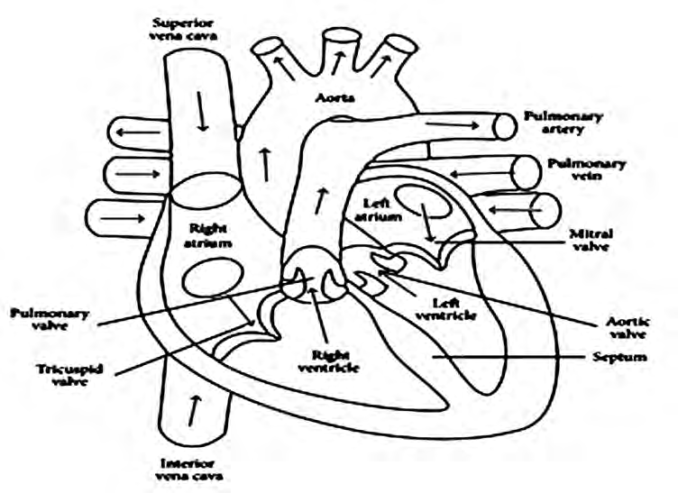
In a heart diagram, right and left mean the right and left of the person whose heart it is, not yours as the reader. So the right atrium appears on your left and the left ventricle on your right, just as a friend standing face to face with you has their right hand on your left side.
The heart has four valves: the tricuspid valve, the bicuspid valve, the pulmonary valve and the aortic valve. A valve works like a one-way door. It opens to let blood go forward and shuts so that the blood cannot come back.
Valve: a door-like flap found between the chambers of the heart and at the openings of the arteries that leave it. Its job is to stop blood flowing in the wrong direction.
| Valve | Where it sits |
| Tricuspid valve | Between the right atrium and the right ventricle |
| Bicuspid valve, also called the mitral valve | Between the left atrium and the left ventricle |
| Pulmonary valve | Where the pulmonary artery leaves the right ventricle |
| Aortic valve | Where the aorta leaves the left ventricle |
The valve marked Mitral valve in the diagram is the bicuspid valve. It has two flaps, which is where the name bicuspid comes from. The tricuspid valve has three flaps.
Functions of the Heart
Day and night, without stopping, the heart does three main jobs:
- It sends blood to every organ of the body.
- It sends the impure, carbon dioxide-rich blood that returns from the organs to the lungs.
- It collects the pure, oxygen-rich blood that comes back from the lungs and sends it out to every organ of the body.
The Blood Circulation Process
The heart is the most important organ of circulation. It works like a pump: when it squeezes, or contracts, it pushes blood out, and when it relaxes it fills with blood again. One full journey of the blood, from the body to the heart, on to the lungs to be cleaned, back to the heart and out to the body again, happens in this order:
- Impure, carbon dioxide-rich blood from the cells, tissues and organs of the body flows through the superior vena cava and the inferior vena cava and collects in the right atrium.
- When the right atrium is full, it contracts. The tricuspid valve opens and the blood passes into the right ventricle.
- When the right ventricle is full, it contracts. The pulmonary valve opens and the impure blood travels along the pulmonary artery to the lungs.
- In the tiny air sacs of the lungs, called alveoli, carbon dioxide and oxygen are exchanged. The blood gives up its carbon dioxide there and takes in oxygen, and so it becomes pure.
- Four pulmonary veins bring this pure blood from both lungs to the left atrium.
- When the left atrium is full of pure blood, it contracts. The bicuspid valve opens and the pure blood passes into the left ventricle.
- When the left ventricle is full, it contracts. The aortic valve opens and the pure blood enters the aorta.
- The aorta carries the pure blood to every part of the body. Once the body has used its oxygen, the blood is impure again and the whole journey starts over.
In one complete round, blood passes through the heart twice: once through the right side on its way to the lungs, and once through the left side on its way to the body. That is why circulation in the human body is studied as two separate kinds.
Systemic Circulation
Systemic circulation: the circulation that carries pure, oxygen-rich blood from the heart through the aorta to the cells and tissues of the body, and brings the carbon dioxide-rich blood produced there back to the heart through the superior and inferior venae cavae.
In systemic circulation, blood carrying oxygen and nutrients leaves the left ventricle through the aorta. It flows along the branches and smaller branches of the arteries until it reaches the capillaries in every part of the body. Inside the cells, oxygen is used to release energy from food. This is called oxidation. The carbon dioxide made by oxidation mixes into the blood. The blood, now impure, flows from the capillaries into the small branches of the veins, then into larger branches, and finally into the two great veins, the superior vena cava and the inferior vena cava, which empty it into the right atrium.
Pulmonary Circulation
Pulmonary circulation: the process that carries impure, carbon dioxide-rich blood from the heart to the lungs, and brings oxygen-rich blood from the lungs back to the heart.
In pulmonary circulation, blood leaves the right ventricle through the pulmonary valve and travels along the right and left pulmonary arteries to the air sacs of the right and left lungs. There it gives up its carbon dioxide. It picks up oxygen from the air in the air sacs and returns through the pulmonary veins to the left atrium. From there, systemic circulation takes over again. In this way blood keeps circulating through our body steadily, without a break.
Systemic and Pulmonary Circulation Compared
| Systemic circulation | Pulmonary circulation |
| Starts from the left ventricle. | Starts from the right ventricle. |
| Blood leaves the heart through the aorta. | Blood leaves the heart through the pulmonary artery. |
| Pure blood travels to the cells and tissues of the body. | Impure blood travels to the air sacs of the lungs. |
| In the cells, blood gives up oxygen and takes in carbon dioxide. | In the lungs, blood gives up carbon dioxide and takes in oxygen. |
| Impure blood returns to the right atrium through the venae cavae. | Pure blood returns to the left atrium through the pulmonary veins. |
| Its arteries carry pure blood and its veins carry impure blood. | Its artery carries impure blood and its veins carry pure blood. |
The full lesson is waiting for you
The notes are free to read in full. A free account opens the rest of the practice, and tracks your progress and confidence score for every subject. Presentations and videos need approval from Learn Yeti.