Two Groups of Female Reproductive Organs
In a woman's body, several organs work together so that a baby can be conceived, can grow and can be born. Together these organs make up the female reproductive system. To study them clearly, we divide them into two main groups. The external organs are the ones on the outside of the body. The internal organs lie protected inside the lower part of the abdomen. Besides these, the breast is counted as an accessory organ, because it provides milk for the baby after birth.
- External organs: the parts on the outside of the body, known together as the vulva.
- Internal organs: the vagina, the uterus, the fallopian tubes and the ovaries, all inside the body.
- Accessory organ: the breast, which makes milk for the baby.
Knowing the correct names of the body's parts is nothing to be shy about. With the right names, it is easier to look after your body and to tell a health worker clearly when something feels wrong.
External Organs: The Vulva
The female reproductive organs that can be seen on the outside of the body, between the thighs, are together called the vulva. The vulva is not one single organ. It is made of several parts, and they are easiest to learn in order from top to bottom.
Vulva: the name given to all the external female reproductive organs taken together.
Mons pubis: the thick pad of skin and fat at the very front of the vulva. Once a girl reaches puberty, it becomes covered with hair.
Labia majora (outer lips): the two thick folds of fatty skin on either side of the vulva. They contain many oil glands and sweat glands. The short hair that grows around them is called pubic hair.
Labia minora (inner lips): the thinner folds of skin lying inside the labia majora, one on each side. The clitoris sits above them, and between them are the vaginal opening and the urethral opening.
Clitoris: a small organ just above the point where the labia minora join. It is a woman's most sensitive external sex organ. It is similar to the male penis, because it is made of the same kind of erectile tissue, tissue that can become firm and upright.
Hymen: a thin membrane that partly covers the opening of the vagina. In a girl who has not had sexual intercourse, part of it breaks at her first menstruation. It breaks completely after sexual intercourse and after childbirth. It also commonly tears during ordinary activities such as riding a bicycle, climbing a tree or running in a race.
The hymen can tear during everyday activities such as cycling, climbing or running. So the state of the hymen can never be used to judge a girl's character.
Perineum: the area that begins at the lower edge of the vulva and stretches back to the anus. It is made of connective tissue, muscle and fat.
Taken in order from top to bottom, the mons pubis comes first. The labia majora lie on both sides, with the labia minora inside them. Where the labia minora meet at the top sits the clitoris, covered by a small hood of skin called the prepuce. The space between the labia minora is called the vestibule, and it holds two openings. The small upper one is the urethral opening, through which urine leaves the body. The larger one below it is the vaginal opening. Below these lie the perineum and, last of all, the anus.
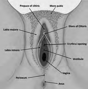
Urine and menstrual blood do not leave through the same opening. Urine comes out through the urethral opening, while menstrual blood flows from the uterus down the vagina and out through the vaginal opening. The anus is a third, separate opening, below the perineum.
Internal Organs
Further inside, protected within the lower part of the abdomen, lie the internal reproductive organs. There are four of them:
- the vagina
- the uterus
- the fallopian tubes
- the ovaries
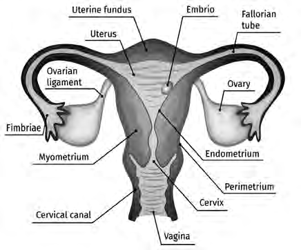
These organs are joined to one another. The uterus sits in the middle. From each side of its upper part, a fallopian tube reaches out towards an ovary. The narrow lower end of the uterus opens into the vagina, and the vagina opens to the outside at the vulva. In this way the organs form one connected passage from the ovaries to the outside of the body.
The Vagina
Vagina: a soft, muscular tube that can stretch and shrink back like a balloon, joining the external reproductive organs to the internal ones.
The vagina lies between the thighs, with the urinary bladder in front of it and the rectum behind it. It is about 15 cm long and 3 to 6 cm wide. Because it links the outer organs with the inner ones, it works like a bridge between the two. Its walls can stretch a great deal when a baby is born and then shrink back again. The vagina has these functions:
- It receives the man's penis during sexual intercourse.
- It receives the sperm during sexual intercourse.
- It forms the birth canal, the passage through which a baby is born.
- It lets menstrual blood from the uterus flow out of the body.
The Uterus
Uterus (womb): a hollow, pear-shaped organ made of smooth muscle tissue, in which a baby lives and grows before it is born.
The uterus is wide at the top and narrow at the bottom, so it looks like an upside-down pear. Its upper part is joined to the fallopian tubes on each side, and its lower part is joined to the vagina. It is a sac that can stretch, so as a baby grows inside it, the uterus grows larger too. Normally it weighs only about 30 to 40 grams.
The uterus is divided into three parts:
- Fundus: the rounded top part of the uterus
- Body (corpus): the main middle part
- Cervix: the narrow lowest part, the neck of the uterus, which opens into the vagina
The wall of the uterus is made of three muscular layers. From the outside in, they are:
- Perimetrium: the outermost layer
- Myometrium: the middle layer
- Endometrium: the innermost layer, which lines the inside of the uterus
When a fertilized egg reaches the uterus, it attaches to the inner surface. There its cells divide again and again, and it develops into a foetus. The foetus stays in the uterus for nine months until it is fully developed, and then the baby is born. The functions of the uterus are:
- It keeps the menstrual cycle regular.
- It prepares a suitable environment for the foetus.
- It receives the fertilized egg and gives it nutrients and protection.
- It lets the foetus grow and develop, and helps the baby to be born after nine months.
The Fallopian Tubes
Fallopian tube: one of the two tubes that spread out like arms from either side of the upper uterus and carry an egg from the ovary to the uterus.
The fallopian tube is also called the uterine tube. At the outer end of each tube are finger-like parts called fimbriae. The fimbriae spread out close to the ovary, so they are ready to catch the egg as soon as the ovary releases it and guide it into the tube.
The main job of the tube is to receive the egg from the ovary and carry it to the uterus. Fertilization also usually takes place inside this tube. The fertilized egg then moves on into the uterus and settles there.
Fertilization: the joining of a sperm with an egg, which usually happens inside the fallopian tube.
The Ovaries
Ovaries: the two organs, one on each side of the uterus, that release eggs and produce the hormones oestrogen and progesterone.
Each ovary holds many immature cells called Graafian follicles, or ovarian follicles. Once a girl reaches puberty, one of these follicles ripens every month and an egg comes out. It happens in this order:
- One follicle ripens.
- The ripe follicle bursts and releases one egg.
- The fimbriae catch the egg.
- The egg moves along the fallopian tube towards the uterus.
As well as releasing eggs, the ovaries produce two hormones, oestrogen and progesterone. These same hormones also make a girl's breasts grow.
An Accessory Organ: The Breast
Breast: an accessory gland of the female reproductive system that produces milk after a baby is born.
The breast does not take part in conceiving or carrying a baby, but it makes the milk the baby needs once it is born. That is why it is called an accessory organ. Breasts begin to develop when a girl reaches puberty. They grow once the ovaries start producing oestrogen and progesterone, and during pregnancy the production of these hormones becomes even stronger. After the baby is born, the pituitary gland produces a hormone called prolactin, which helps the breasts make milk.
Each breast is made of fatty tissue together with fibrous tissue. In the very centre of the breast is a dark tip called the nipple. The milk made inside the breast comes out through the nipple.
The Organs at a Glance
| Organ | Group | Key fact |
| Mons pubis | External | Pad of skin and fat, covered with hair after puberty |
| Labia majora | External | Thick fatty folds with oil and sweat glands |
| Labia minora | External | Thin inner folds around the vaginal and urethral openings |
| Clitoris | External | Very sensitive, made of erectile tissue |
| Hymen | External | Thin membrane partly covering the vaginal opening |
| Perineum | External | From the lower vulva to the anus |
| Vagina | Internal | Muscular tube about 15 cm long, the birth canal |
| Uterus | Internal | Pear shaped, 30 to 40 grams, where the baby grows for nine months |
| Fallopian tubes | Internal | Carry the egg to the uterus, where fertilization usually happens on the way |
| Ovaries | Internal | Release one egg a month, make oestrogen and progesterone |
| Breast | Accessory | Makes milk for the baby, which leaves through the nipple |
The full lesson is waiting for you
The notes are free to read in full. A free account opens the rest of the practice, and tracks your progress and confidence score for every subject. Presentations and videos need approval from Learn Yeti.