What Is an Animal Tissue?
An animal's body is built from millions of cells, and those cells do not sit scattered at random. Cells that do the same kind of work join together into a group, and that group carries out one definite job for the body. Blood transports materials, muscles produce movement, nerves carry messages. Each of these jobs belongs to a different tissue.
An animal tissue is a group of animal cells joined together, and every animal tissue performs one definite function.
Animal tissues are generally of four kinds:
- Epithelial tissue, the covering and lining tissue
- Muscular tissue, the tissue of movement
- Connective tissue, the joining and supporting tissue
- Nervous tissue, the message carrying tissue
Epithelial Tissue: The Body's Lining
Epithelial tissue is the group of cells that forms the outermost layer of an animal's body or of its organs.
Epithelial tissue may be made of one kind of cell or of several kinds together. Its cells stay fixed on a thin base called the basement membrane. The main jobs of epithelial tissue are covering surfaces, protecting the parts underneath, secreting useful substances, excreting wastes and absorbing materials.
On the basis of the nature and thickness of the cells, epithelium is divided into several kinds. The four important ones are pavement epithelium, cubical epithelium, columnar epithelium and glandular epithelium.
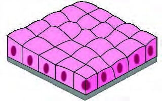
In pavement epithelium, flat plate like cells with many sides sit fitted tightly against each other, the way tiles fit on a floor. There is only a single layer of cells. It is also called squamous epithelium. Its work is covering, protecting and filtering, and it forms the outer covering of organs such as the heart, the lungs and the kidneys.
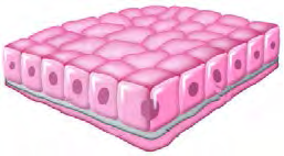
Cubical epithelium is a single layer of cube shaped cells. Tissue of this kind builds the small tubes inside the kidneys, the thyroid gland and the various ducts of excretory glands, the inner part of the uterus and the air passages. Its work is covering, protecting and secreting.
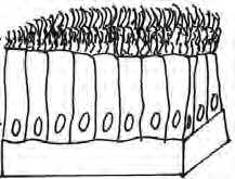
Columnar epithelium is made of tall, pillar shaped cells standing on the basement membrane in a single layer. Their free surface may be covered with tiny hair like structures called cilia, which keep the nearby fluid moving to and fro. This tissue is found in secreting organs such as the gall bladder and the salivary glands, and its main work is secretion and absorption.
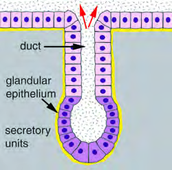
The glands of the body are lined by secretory epithelial cells, and the tissue built from such cells is called glandular epithelium. These cells produce substances the body needs, such as hormones, enzymes, mucus, saliva and digestive juices.
| Epithelium | Cell shape | Where it is found | Main work |
| Pavement (squamous) | Flat, plate like | Outer covering of the heart, lungs and kidneys | Covering, protecting, filtering |
| Cubical | Cube shaped | Kidney tubules, thyroid and excretory gland ducts, inner uterus, air passages | Covering, protecting, secreting |
| Columnar | Tall, pillar like; may carry cilia | Gall bladder, salivary glands and other secreting organs | Secretion and absorption |
| Glandular | Secretory cells | Glands of the body | Making hormones, enzymes, mucus, saliva, digestive juices |
Muscular Tissue: The Movers of the Body
Muscular tissue is the tissue that produces movements such as contraction and relaxation in different parts of an animal's body. It is built from groups of long, thread like muscle fibre cells.
The main function of muscular tissue is to bring movement to the body. It is of three types: skeletal muscular tissue, cardiac muscular tissue and smooth muscular tissue.
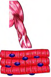
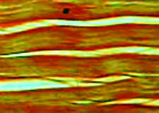
Skeletal muscular tissue has long fibres and stays attached to the bones. Working together with connective tissue, that is with bone, it produces movement in the body, which is why it is called skeletal muscle. Dark and light bands are seen across its fibres, so it is also called striped muscle. You can move it whenever you wish, so it is also called voluntary muscle. Lifting your hand and moving your legs are its work.
Cardiac muscular tissue is found only in the walls of the heart. It contracts and relaxes without ever stopping, and this contraction and relaxation creates the heartbeat, by which the heart pumps blood. Bands are seen in it too, but this muscle cannot be moved at will.
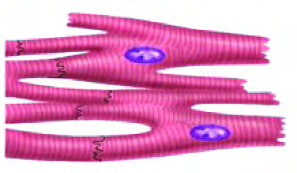
Do not think every banded muscle is voluntary. Cardiac muscle shows bands, yet it cannot be moved at will. Judging a muscle voluntary just because it is striped is a mistake.
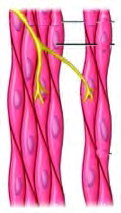
Smooth muscular tissue is made of groups of spindle shaped muscle fibres. Its cells show no bands, which is why it looks smooth. Tissue of this kind is found in the inner parts of the stomach and the intestines, and also in organs such as the urinary tract and the blood vessels. It too cannot be moved at will, so it is called involuntary muscle.
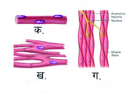
| Muscular tissue | Bands seen? | Moves at will? | Where it is found |
| Skeletal | Yes | Yes (voluntary) | Attached to the bones |
| Cardiac | Yes | No | Only in the walls of the heart |
| Smooth | No | No (involuntary) | Stomach, intestines, urinary tract, blood vessels |
Connective Tissue: The Body's Binder
Connective tissue is the tissue that joins the different tissues and organs found in the body.
The cells of connective tissue stay separate from one another and are spread through every part of the body. Its main work is to support the body and to keep the cells and tissues connected with each other. Between its cells there are gaps called intercellular spaces, and these spaces stay filled with solid or liquid material.
The solid or liquid material that fills the intercellular spaces of connective tissue is called the matrix. It contains water, fibres and minerals.
On the basis of its form, connective tissue is divided into three types:
- Soft connective tissue: fibrous and adipose
- Hard connective tissue: bone and cartilage
- Fluid connective tissue: blood and lymph
Soft Connective Tissue
In soft connective tissue the cells are loosely arranged, and the intercellular spaces hold plenty of fibres and matrix. In some of these tissues the cells store a large amount of fat. Soft connective tissue is of two kinds: fibrous and adipose.
Fibrous connective tissue is built from fibres joined either loosely or densely. The loosely joined kind is found under the skin, between muscles and around blood vessels. Dense fibrous tissue is the most common connective tissue in the body. The densely joined kind can stretch, and it binds organs together and gives them shape. Such tissue is found around the kidneys, the liver, the glands and the muscles, and it also carries out repair work.
A tendon is the fibrous connective tissue that joins a muscle to a bone.
A ligament is the fibrous connective tissue that joins one bone to another bone.
Many students swap tendon and ligament in the exam. A tendon ties muscle to bone, while a ligament ties bone to bone. Be sure which pair each word joins before you write it.
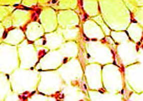
Adipose connective tissue contains only a few fibres, and its matrix is made of fat or of other soft material. Areolar and adipose tissue are its examples. Areolar tissue lies under the skin and joins the skin to the muscles. Adipose tissue is fat itself. Inside the body it works as a cushion, and it plays an important part in controlling body temperature.
Hard Connective Tissue: Bone and Cartilage
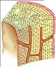
Bone and cartilage belong to hard connective tissue. The cell found in these tissues is called an osteocyte. From outside, a bone is covered by a thin membrane called the periosteum. Calcium stays deposited in bones, which is what makes them so hard. The bones of the skull, the limbs and the backbone are made of hard bone. These connective tissues join and support the different tissues of the body.
Cartilage is found in the nose, the ears, the throat, between the bones of the backbone and at the ends of long bones. It reduces friction in the moving parts of the body and protects them from shock. The cartilage formed in infancy slowly hardens as calcium salts are laid down in it.
Fluid Connective Tissue: Blood and Lymph
Some connective tissues exist in a fluid state. They act as connectors between the different systems of the body and help with transport inside it. Animals have two kinds of fluid connective tissue: blood and lymph.
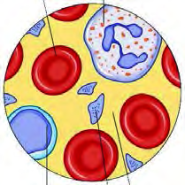
Blood is the red fluid connective tissue of the body. Its work is to carry different substances from one part of the body to another.
Blood is 55 percent plasma and 45 percent blood cells. Plasma itself is 90 percent water and 10 percent solid material. Nutrients, enzymes, hormones and other substances dissolve in the plasma and travel in it from one part of the body to another. Blood contains three kinds of cells:
- Red blood cells: they carry haemoglobin, with whose help blood picks up oxygen from the lungs and delivers it everywhere, then collects carbon dioxide from the body's parts and brings it back to the lungs.
- White blood cells: they destroy harmful germs.
- Platelets: at an injured spot they help the blood to clot, so bleeding stops.
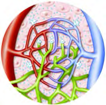
Lymph is the colourless fluid connective tissue found in vessels. It contains no haemoglobin.
Lymph contains plasma and lymphocytes. In the parts of the body that blood itself does not reach, the fluid that filters out of the blood vessels into the intercellular spaces for the tissues is lymph. It nourishes the tissues, protects them and carries away their wastes.
Nervous Tissue and the Neuron
Nervous tissue is the tissue that directs the different organs of an animal's body, carries messages between them and keeps their work coordinated.
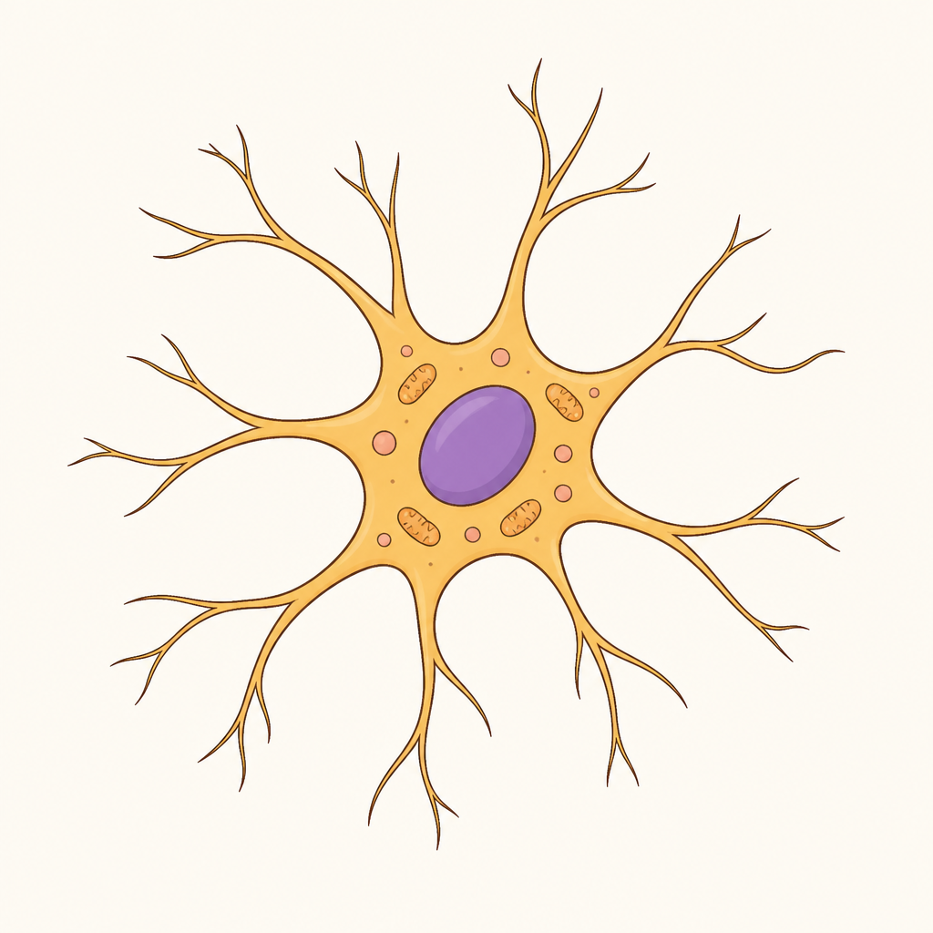
The smallest unit of nervous tissue is the neuron. Nervous tissue is built of neurons, and a connective tissue called neuroglia holds them bound together.
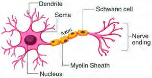
The smallest unit of the nervous system is called a neuron, or nerve cell.
The central cell of a neuron is called the cell body, and it is grey in colour. The single long branch that comes out of it is called the axon, which is white, and the axon may be wrapped in a covering called the myelin sheath. The short branches coming from the cell body are called dendrites. The dendrites bring impulses from other nerve cells to the cell body, while the axon carries the impulse from the cell body onward to other nerve cells.
When your leg gets hurt, the pain of that injury travels all the way to the brain, and only then do you feel it. This is possible because of the communication that neurons carry out among themselves.
The Four Tissues at a Glance
| Tissue | Main work | Examples |
| Epithelial | Covering, protecting, secreting, excreting, absorbing | Covering of heart and lungs, kidney tubules, glands |
| Muscular | Bringing movement to the body | Muscles on the bones, wall of the heart, wall of the intestine |
| Connective | Joining tissues and organs, supporting, transporting | Bone, cartilage, tendon, ligament, blood, lymph |
| Nervous | Directing, communicating, coordinating | Neurons of the brain and nerves |
Each of the four tissues has its own job, yet the body needs all four working together. Even raising your hand uses bone, muscle, tendon, blood and neurons all at once.
The full lesson is waiting for you
The notes are free to read in full. A free account opens the rest of the practice, and tracks your progress and confidence score for every subject. Presentations and videos need approval from Learn Yeti.