How the Body Sends Its Messages
How do we come to know about the changes happening around us? Why does a hand jump back the instant it touches a hot pan? Behind every one of these actions is a communication system inside the body. Our sense organs, nerves and brain work together to gather information about everything around us. The brain controls and operates every organ of the body, and when something in the environment stimulates us, we respond.
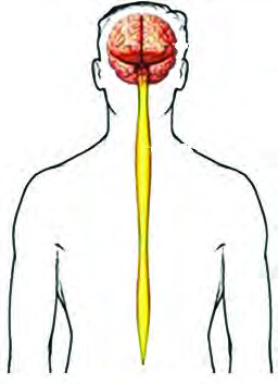
The nervous system is the system formed by a special group of organs that responds to changes in the environment and coordinates the organs of the body. It is made of the brain, the spinal cord and the nerves.
The sense organs are the outer organs of this system, while the brain, the spinal cord and the nerves handle the coordination inside the body. On the basis of its parts and their work, the human nervous system is divided into three parts.
| Division | Main parts | Main job |
| Central nervous system | Brain and spinal cord | Controls and coordinates the whole body |
| Peripheral nervous system | Sensory and motor nerve fibres | Carries messages between the central system and the body |
| Autonomic nervous system | Sympathetic and parasympathetic | Controls the involuntary work of internal organs |
The Central Nervous System
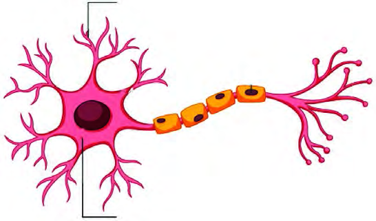
The part of the nervous system that runs down the middle of the body, from the head toward the lower back, is called the central nervous system. It is made of two organs, the brain and the spinal cord. The tissue itself is built of neurons and glia. The neuron is the basic cell of the whole nervous system, and it stays wrapped in a protective covering.
The Brain and Its Protective Layers
The brain is the largest and uppermost part of the central nervous system, kept safe inside the skull. It coordinates and controls every organ of the body. Around the brain lie three protective membranes.
The meninges are the three layered membranes that cover the brain. From the outside inward they are the dura mater, the arachnoid mater and the pia mater.
The dura mater sticks to the inner surface of the skull, and the pia mater sticks to the surface of the brain itself, with the arachnoid mater between them. Between the arachnoid mater and the pia mater there is a space, and that space is filled with a liquid.
Cerebrospinal fluid is the liquid that fills the space between the arachnoid mater and the pia mater. Its main work is to nourish the brain and to protect it, acting as a cushion. It is an alkaline liquid made of water, minerals, glucose and protein.
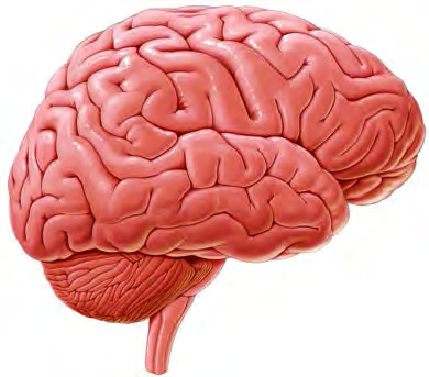
The brain itself has different parts. Its three main parts are the cerebrum, the cerebellum and the medulla oblongata, and each has its own work.
The Cerebrum
The cerebrum is the largest part of the brain, covering about 80% of it. It is divided into a right half and a left half, called cerebral hemispheres, and on the upper, dorsal side a deep fissure separates the two hemispheres. The outer surface of the hemispheres is wrinkled into folds, which increases the surface area. The outer surface of the cerebrum is grey and the inner part is white.
The cerebrum runs and controls many of the body's activities. Alongside that, it does the following work:
- It perceives smell.
- It operates and controls speech, memory and hearing.
- It creates and controls consciousness, thought and excitement.
- It controls the ability to analyse, to put ideas together and to predict.
- It also controls the other parts of the brain.
If the cerebrum is injured, the person may lose consciousness, memory may weaken, and the power of coordination may decline. A serious injury can take a person all the way into a coma.
The Cerebellum
The cerebellum is the second largest part of the brain, covering about 10% of it. It sits behind the cerebrum and above the medulla oblongata as two half rounded parts shaped like a pair of lemon seeds. Like the cerebrum, its outer part is grey matter and its inner part is white matter. Its work is as follows:
- It controls various activities of the body.
- It acts as the coordinator of muscle movement.
- It keeps the body balanced while a person walks.
- It also conducts voluntary movement.
- It maintains muscle tone.
If the cerebellum is injured, the body loses its balance and voluntary movement is affected, which is a state of disability. Alcohol acts on the cerebellum, and that is exactly why a person who has been drinking staggers while walking.
The Medulla Oblongata
The medulla oblongata lies at the lowest part of the brain. It is one of the three parts of the brainstem, and the other two, the pons varolii and the midbrain, serve as the bridge between the spinal cord and the brain. The medulla oblongata itself sits between the spinal cord and the pons varolii. It is shaped like a stick, a narrow tube, and both its outer and inner surfaces are of a whitish grey colour. It passes out of the skull through the opening at its base called the foramen magnum. Its special charge is the control of involuntary actions.
- The medulla oblongata operates breathing.
- It controls vomiting, coughing, sneezing and swallowing.
- It narrows and widens the blood vessels.
- It helps the secretion of hormones, digestive juices and saliva.
- If the medulla oblongata is injured, the person dies immediately.
The Spinal Cord
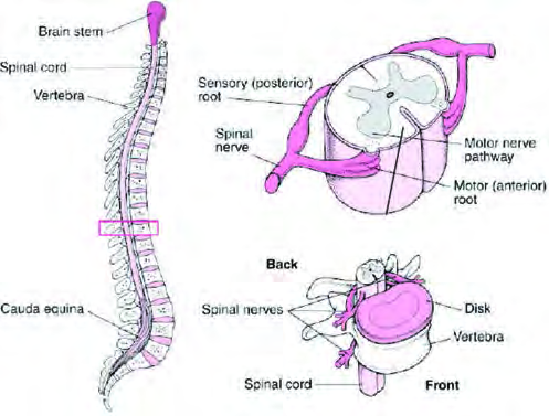
The spinal cord is a long, jelly like structure that runs down inside the backbone. It begins at the base of the medulla oblongata and extends to the second lumbar vertebra of the back. Its outer part is white and its inner part is grey, the opposite arrangement to the cerebrum, and it too is covered by the meninges. It is about 45 cm long and 2 cm in diameter. The spinal cord carries messages between the different parts of the body and the brain, and it also serves as the centre of reflex action.
Nerve Fibres
Nerve fibres are formed when the axons of nerve cells bundle together. The nerves work as messengers for the brain and the spinal cord: some bring news in, others carry orders out. On the basis of their work, nerve fibres are of three types:
- Afferent or sensory nerve: it carries the nerve impulse from a receptor to the brain or the spinal cord.
- Efferent or motor nerve: it carries the message or impulse from the brain or the spinal cord out to the different parts of the body.
- Interneuron: it joins the sensory nerve fibre to the motor nerve fibre and passes the impulse that arrived on the sensory fibre across to the motor fibre.
Many students swap the directions of the sensory and motor nerves. The sensory nerve brings news from the body in to the brain or spinal cord, while the motor nerve takes orders from the brain or spinal cord out to the body. Remember it as senses report in, motors drive out.
The Peripheral Nervous System
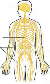
The peripheral nervous system is the part that carries communication between the central nervous system and the different parts of the body. It is built entirely of nerve fibres. These fibres are grouped into two kinds by where they emerge: the nerves that come out of the brain are called cranial nerves, and the nerves that come out of the spinal cord are called spinal nerves.
| Basis | Cranial nerves | Spinal nerves |
| Origin | They emerge from the brain | They emerge from the spinal cord |
| Number | There are 12 pairs | There are 31 pairs |
| Work | They connect the brain with the tongue, eyes, ears, nose and other organs of the head | They bring impulses to the spinal cord from the body and carry them back out |
Reflex Action
Sometimes, when we slip suddenly while walking on the road, the hand presses against the ground before we have even thought about falling. In the same way we snatch a hand back from a hot object, and the eye blinks shut when something is about to enter it. We do many actions of this kind without knowing we are doing them, responding while still unaware of what happened.
A reflex action is the quick, automatic response the body gives when a part of it suddenly comes into contact with something. Reflex actions are conducted by the spinal cord, not decided by the brain.
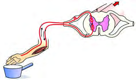
During a reflex action the impulse travels along one fixed path through the nervous system. That path is called the reflex arc.
In the reflex that happens on touching a hot object, the impulse travels through these five stations, in this order:
- Receptor: the nerve ending in the skin that picks up the change in the surroundings.
- Sensory nerve: the nerve that carries the news from the receptor to the spinal cord.
- Connecting nerve: the nerve that brings the impulse down within the spinal cord, coordinating between the sensory nerve and the motor nerve.
- Motor nerve: the nerve that carries the impulse from the spinal cord to the muscles.
- Effector: the muscle that receives the impulse and moves.
In a reflex, the impulse does not have to travel to the brain and back. The spinal cord itself gives the order at once, which is why the response is so fast and the body escapes harm in time.
The Autonomic Nervous System
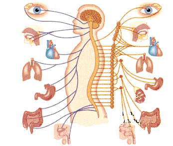
The autonomic nervous system is the part of the nervous system that controls the involuntary work of organs such as the heart, the kidneys and the lungs. Whether a person is awake or asleep, it keeps controlling the muscles and certain glands. It has two parts, the sympathetic nervous system and the parasympathetic nervous system, and the two pull in opposite directions.
| Basis | Sympathetic nervous system | Parasympathetic nervous system |
| Work | It prepares the body to face a sudden emergency | It stops the effects of the sympathetic system |
| Heart and lungs | It speeds up the heart and the lungs | It slows the heartbeat and lowers the blood pressure |
| When it acts | During a sudden event it raises blood pressure and heartbeat so the body produces extra energy, which the organism uses to protect itself | Once the emergency is over, it helps bring the heart and the breathing back to their normal state |
The full lesson is waiting for you
The notes are free to read in full. A free account opens the rest of the practice, and tracks your progress and confidence score for every subject. Presentations and videos need approval from Learn Yeti.