What a Reflected Wave Can Tell
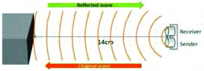
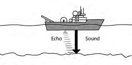
When a wave is sent out, the wave that comes back can tell us what it struck and how far away that thing is. An ultrasonic ruler sends out high frequency sound, times how long the wave takes to bounce off a wall and return, and works out the distance from that time. A fathometer fitted to a ship does the same job at sea. It sends sound towards the sea bed and finds the depth of the water from the time the echo takes to come back. Every one of these instruments rests on a single idea: a reflected wave carries information about the object it struck.
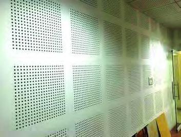
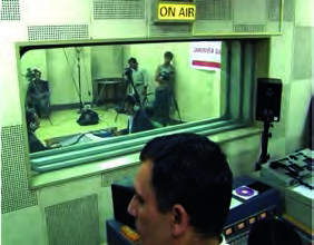
Absorbing sound is another way of putting sound to work. The walls of recording studios and meeting halls are lined with perforated acoustic boards. These boards absorb sound, so no echo can form and outside noise cannot get in.
Doctors use this same reflection principle inside the human body. Three main wave based techniques let them build pictures of the internal organs without cutting the body open: ultrasonography, X-ray imaging and the CT scan.
Ultrasonography
Ultrasonography is the medical technique of examining the internal parts of the body using ultrasound waves. In everyday speech it is called a video X-ray.
This technique uses a device called a transducer. The transducer produces ultrasound of very high frequency, and this ultrasound is aimed into the body. As the waves travel through the body, the different layers inside reflect them back, and the same transducer receives the returning waves. Signals carrying the return time and frequency of these waves go to a computer, and from that data the computer builds up a picture of the structure inside.
The picture of an internal organ that the computer builds from reflected ultrasound data is called a sonogram.
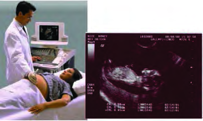
Ultrasound does not harm the body's cells. That is why it is considered the safest technique for check-ups that must be repeated and for examining pregnant women.
What Ultrasound Can Diagnose
Ultrasound gives good images of the body's soft tissues and fluids, so it is used in many different kinds of examination.
- Finding stones in the kidneys, the gall bladder and similar organs
- Detecting tumours in the brain, the pancreas and other organs
- Checking liver disease and problems of the intestines
- Examining problems of the uterus in women
- Checking the size, position and organ movements of a baby growing in the womb, and spotting any abnormal condition
With three dimensional, or 3D, ultrasonic technique the condition of the baby can be observed even more clearly. Ultrasound is also used to examine the beating of the heart, the condition of its valves and how well they are working.
Echocardiography is the technique of examining the movement of the heart and the state and working of its valves using ultrasound.
X-ray Imaging
An X-ray is an electromagnetic wave. Unlike sound it needs no medium, and it can pass right through many parts of the body.
X-ray imaging is the technique of examining the body by preparing pictures of the internal organs using X-rays. In ordinary speech, having X-ray photography done is simply called getting an X-ray.
X-ray photography obtains an X-ray photo from the shadows that the internal organs form when X-rays are sent through the body. In the traditional technique, the person is placed at a fixed distance in front of the X-ray machine. When the machine is switched on, the X-rays that pass through the person's body fall on an X-ray film placed on the other side and form a shadow there. From that shadow, the X-ray photo is prepared from the film. On the photo, the parts in shadow appear white and the rest appears black. In the modern technique an electronic recording device is used instead of photographic film, so the shadows of the internal organs can be observed directly on a computer screen.
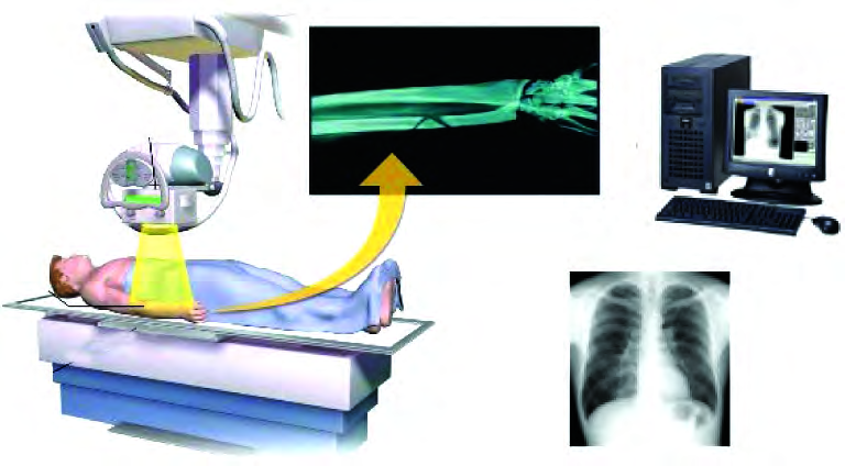
Reading an X-ray Photo
The bones in our body absorb more of the X-rays, so on an X-ray photo the shadow of bone appears dense white. Other tissues, such as muscle and fat, let most of the X-rays pass through and absorb only a little, so these parts show only a very faint shadow. Parts of the body that hold air, such as the lungs, let X-rays through easily, so they appear black on the photo. X-rays are used to check broken or cracked bones, the roots of teeth, infections, tumours, blockages in blood vessels and similar problems.
High energy X-rays are radiation that is harmful to health. Too much X-ray exposure can destroy the body's cells and cause defects in the genes, which raises the risk of cancer. While the X-ray machine is switched on, X-rays are scattered around the room, so an apron of lead must be worn to block the scattered rays. During check-ups, the body must not be exposed to X-rays for a long time or again and again.
The physicist Wilhelm Roentgen discovered X-rays in 1895, while passing an electric current through gases kept at low pressure in his laboratory.
The CT Scan
The Computerized Tomography scan, or CT scan, is a technique developed from X-ray photography, in which a computer and an X-ray machine together build two dimensional and three dimensional pictures of the body.
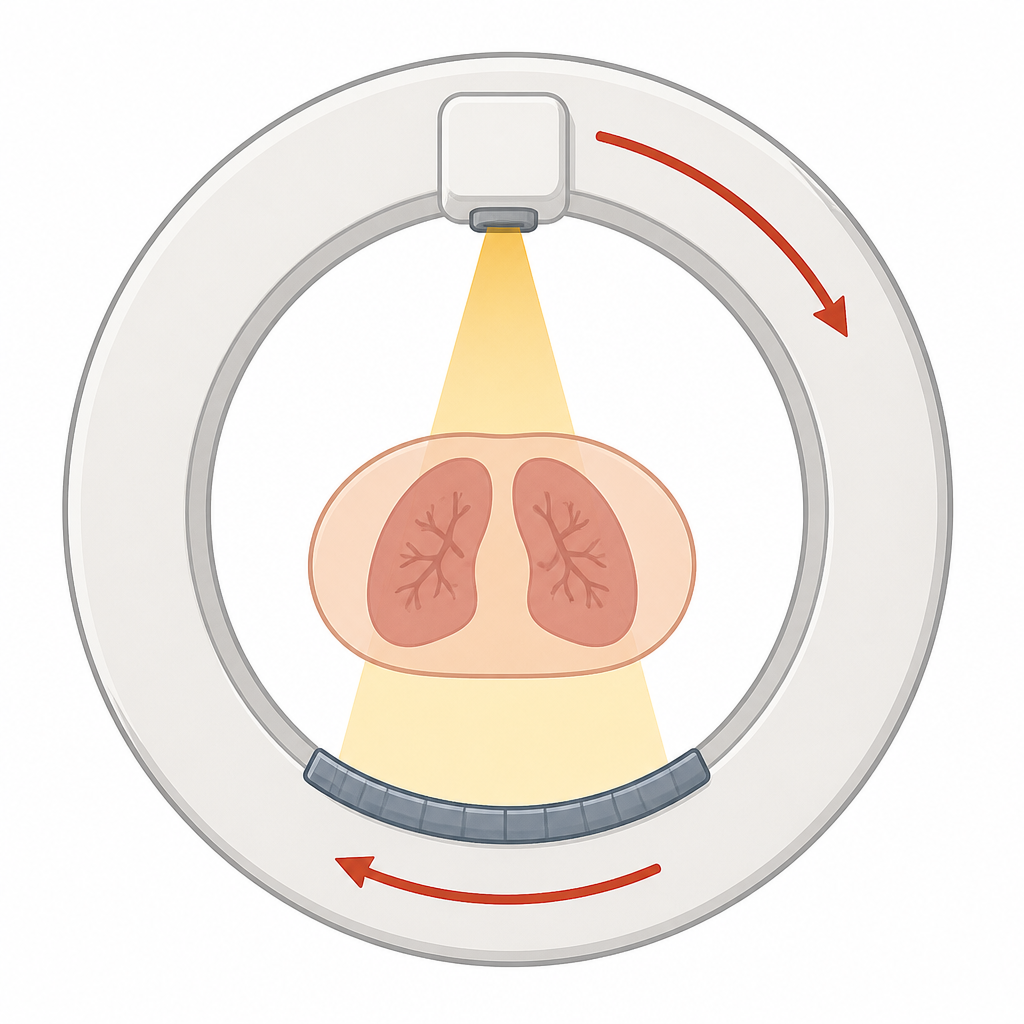
For a CT scan, the person is passed into a doughnut shaped machine. The X-ray source inside the machine can travel right around it, through a full angle of 360°. Directly opposite the source sits the X-ray detector, and the machine is built so that as the source turns, the detector turns with it.
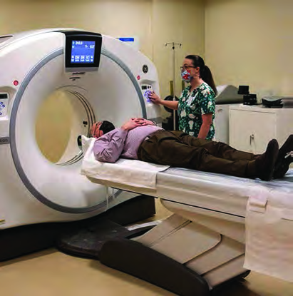
During the scan, a powerful narrow X-ray beam from the source is aimed at the part of the body that holds the organ to be examined. As the source is swung around the body, it is aimed at the same slice of the body from a different direction at every interval of 45°, and the data is collected through the computer. The computer processes this data and prepares two dimensional, or 2D, cross sectional pictures, called tomographic images. For a detailed picture of the structures inside, the data from the X-ray slices is combined to build a three dimensional, or 3D, image.
What Makes a CT Scan Special
A tomographic image holds far more detailed information than a traditional X-ray photo. Even the state of the soft tissues inside the body can be observed in it. The 3D image makes it easy to identify the parts inside the body and to pick out any change or abnormal condition in the internal structures. CT scans are used to examine injuries inside the body, clotted blood, tumours, the condition of the brain, the condition of the lungs and similar problems.
The Three Methods Compared
| Technique | Wave used | How the picture forms | Main uses |
| Ultrasonography | Ultrasound (a longitudinal sound wave) | The computer builds a sonogram from reflected wave data | Stones, tumours, the uterus, the baby in the womb, the heart |
| X-ray photography | X-rays (an electromagnetic wave) | Rays passing through the body cast a shadow on a film or detector | Broken bones, roots of teeth, infections, blockages |
| CT scan | X-rays (an electromagnetic wave) | The computer joins slices taken by a rotating source into 2D and 3D images | Internal injuries, blood clots, detailed checks of the brain and lungs |
Which technique to choose depends on what has to be examined. A baby in the womb is checked with ultrasonography, because X-rays are harmful radiation while ultrasound does not harm cells. For a broken bone, an ordinary X-ray is enough. When a detailed three dimensional examination of an organ such as the brain is needed, a CT scan is done.
The full lesson is waiting for you
The notes are free to read in full. A free account opens the rest of the practice, and tracks your progress and confidence score for every subject. Presentations and videos need approval from Learn Yeti.