The Heart: Centre of the Circulatory System
Every cell of your body needs oxygen and digested food all the time, and every cell makes waste that has to be taken away. Blood carries all of these, but blood cannot move by itself. It has to be pushed through a long network of blood vessels, without a break, every minute of your life. The organ that does this pushing is the heart. The heart is the central organ of the circulatory system. It pumps blood to each cell and tissue of the body and receives the blood that returns from them.
Heart: a triangular or conical muscular organ made of cardiac muscles, which contracts and expands regularly to pump blood throughout the body.
The heart is made of cardiac muscle, a kind of muscle found only in the heart. The muscles of your arm get tired after carrying a heavy bucket of water for a while, but cardiac muscle keeps contracting and relaxing regularly throughout life. Each contraction squeezes blood out of the heart into the blood vessels. Each relaxation lets the heart fill with blood again. This steady squeeze and relax is what you feel as your heartbeat.
The heart of a goat or a buffalo is built on the same plan as the human heart. It has the same four chambers and the same large blood vessels at its top, and differs only in size. A fresh one from a butcher's shop is therefore a good real specimen for seeing the outer parts of a heart and, once it is cut lengthwise, its inner parts.
Wear gloves while touching and handling an animal heart, and wash your hands with soap and water when you finish. Cut it only when your teacher is present. Use the scissors and scalpel from the dissection box carefully, and never point the blade towards yourself or anyone else.
Where the Heart Lies
The heart sits inside the thoracic cavity, the space in the chest enclosed by the ribs. There it lies between the two lungs, with the breastbone in front of it and the backbone behind it. The heart is not exactly in the middle. It is slightly tilted towards the left side, and about two-thirds of it lies to the left of the thoracic cavity. This is why the beat is felt most clearly when you place your palm on the chest just to the left of the centre.
The Heart's Protective Cover: Pericardium
Because the heart never rests and is so important, it is well protected. It is enclosed within a double-layered protective membrane called the pericardium. The space between the two layers is filled with a fluid called pericardial fluid. This fluid works like a cushion. Just as the soft padding inside a helmet protects the head from a knock, pericardial fluid protects the heart from external shocks and injuries.
Pericardium: the double-layered protective membrane that encloses the heart.
Pericardial fluid: the fluid filling the space between the two layers of the pericardium, which protects the heart from external shocks and injuries.
Shape, Size and Weight of the Heart
The human heart is triangular or conical in shape. It is broad at the top and narrows towards a pointed tip at the bottom. Its size is about the size of its owner's closed fist, so a child's heart is smaller than an adult's and grows as the body grows. On average the human heart weighs about 300 grams, roughly the weight of two medium-sized apples.
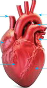
Looking at the front of the heart from outside, these parts can be seen:
- Right auricle: the upper chamber on the heart's right side
- Left auricle: the upper chamber on the heart's left side
- Right ventricle: the lower chamber on the heart's right side
- Left ventricle: the lower and strongest chamber on the heart's left side, which forms the pointed tip
- Superior vena cava: the large vein joining the top of the heart on its right side
- Aorta: the thick artery that arches out from the top of the heart
- Pulmonary vein: the vessels from the lungs that join the heart on its left side
Right and left in a heart always mean the right and left of the person the heart belongs to. In a front view the heart faces you, so its right side appears on your left in the picture. It is the same as a friend facing you, whose right hand is on your left.
The Heart's Own Blood Supply
The heart is a muscle that works without stopping, so its own tissue needs a large supply of oxygen and food. The blood flowing through its chambers does not feed the thick heart wall directly. Instead, a hair-like, very fine network of capillaries covers the surface of the heart. The coronary artery supplies oxygenated blood to the heart muscle. After the muscle has used the oxygen, the coronary sinus vein carries the deoxygenated blood to the right auricle of the heart.
Coronary artery: the artery that supplies oxygenated blood to the heart muscle itself.
Coronary sinus vein: the vein that carries deoxygenated blood from the heart muscle to the right auricle.
Four Chambers and the Septum
Inside, the human heart is hollow and divided into four chambers, so it is called four-chambered. The upper two chambers are called auricles or atria. One of them is an atrium, and the two together are atria. The lower two chambers are called ventricles. Down the middle of the heart runs a thick muscular wall called the septum. The septum divides the heart into a right part and a left part. The right part carries deoxygenated blood and the left part carries oxygenated blood, and because of the septum these two kinds of blood never mix inside the heart.
So the four chambers are named the right auricle, right ventricle, left auricle and left ventricle. Blood vessels are joined to these chambers, and through them the heart receives and supplies blood. The auricles receive blood coming from various parts of the body. The ventricles send blood out to various parts of the body.
Auricle (atrium): an upper chamber of the heart, which receives blood arriving from the body or the lungs.
Ventricle: a lower chamber of the heart, which pumps blood out of the heart to the body or the lungs.
Septum: the thick muscular wall in the middle of the heart that divides it into right and left parts and prevents oxygenated and deoxygenated blood from mixing.
Auricles receive blood. Ventricles send blood out.
Why the Ventricle Walls Are Thicker
The heart pumps blood to different parts of the body with great pressure. An auricle only has to push blood a short distance, down into the ventricle just below it. A ventricle has to push blood out of the heart and along the blood vessels. To withstand and produce this pressure, the walls of the ventricles are thicker than the walls of the auricles.
The two ventricles are not equal either. The right ventricle pumps blood only up to the lungs, which are right beside the heart. The left ventricle has to pump blood with great pressure to every organ of the body, from the head down to the toes. Therefore the wall of the left ventricle is thicker than the wall of the right ventricle. Think of two people watering with a pipe. One waters a flower pot a step away, while the other must send water to the far end of a long field. The second needs far more force.
| Chamber | Receives blood from | Sends blood to | Wall |
| Right auricle | Body (superior and inferior vena cava) and coronary sinus vein | Right ventricle | Thin |
| Right ventricle | Right auricle | Lungs, through the pulmonary artery | Thick |
| Left auricle | Lungs, through the pulmonary vein | Left ventricle | Thin |
| Left ventricle | Left auricle | All organs of the body, through the aorta | Thickest |
Blood Vessels Joined to the Heart
Five main blood vessels are joined to the chambers of the heart. Two of them bring deoxygenated, or impure, blood from the body. One takes that blood to the lungs. One brings oxygenated, or pure, blood back from the lungs. One sends the pure blood out to the whole body.
- Superior vena cava: brings impure blood from the upper parts of the body to the right auricle.
- Inferior vena cava: brings impure blood from the lower parts of the body to the right auricle.
- Pulmonary artery: carries deoxygenated blood from the right ventricle to the lungs for oxygenation.
- Pulmonary vein: carries oxygenated blood from the lungs to the left auricle.
- Aorta: carries oxygenated blood from the left ventricle to the various parts of the body.
Vena cava: the large veins that bring impure blood from the body to the right auricle. The one from the upper body is the superior vena cava and the one from the lower body is the inferior vena cava.
Aorta: the largest artery of the body, leaving the left ventricle and carrying pure blood to the whole body.
Arteries carry blood away from the heart and veins carry blood towards it. Most arteries carry oxygenated blood and most veins carry deoxygenated blood, but there are two exceptions. The pulmonary artery is the only artery that carries deoxygenated blood, and the pulmonary vein is the only vein that carries oxygenated blood. A vessel is named artery or vein by the direction its blood flows, not by whether the blood is pure or impure.
The Four Valves of the Heart
Blood inside the heart must always move forward: from auricle to ventricle, and from ventricle out into the arteries. It must never flow back. This is the job of the valves. A valve is a flap of tissue that opens to let blood pass one way and closes when blood tries to come back, like a door that swings open in one direction only. There are four valves in the heart.
Tricuspid valve: the valve between the right auricle and the right ventricle. When it opens, blood passes from the right auricle to the right ventricle.
Bicuspid valve or mitral valve: the valve between the left auricle and the left ventricle. When it opens, pure blood flows from the left auricle to the left ventricle.
Pulmonary valve (also called the pulmonic valve): the valve between the right ventricle and the pulmonary artery.
Aortic valve: the valve between the left ventricle and the aorta.
| Valve | Where it is | When it opens |
| Tricuspid | Between right auricle and right ventricle | When the auricles contract |
| Bicuspid (mitral) | Between left auricle and left ventricle | When the auricles contract |
| Pulmonary | Between right ventricle and pulmonary artery | When the right ventricle contracts |
| Aortic | Between left ventricle and aorta | When the left ventricle contracts |
Valves let blood flow in one direction only and stop it from flowing back.
How Blood Passes Through the Heart
Following one drop of impure blood that has come back from the body, it moves through the heart in this order:
- Impure blood from the upper body comes through the superior vena cava, and from the lower body through the inferior vena cava, into the right auricle.
- When the auricles contract, the tricuspid valve opens and the blood passes from the right auricle into the right ventricle.
- When the right ventricle contracts, the tricuspid valve closes and the pulmonary valve opens. The deoxygenated blood is pumped through the pulmonary artery to the lungs for oxygenation.
- The blood, now oxygenated in the lungs, returns through the pulmonary vein to the left auricle.
- When the auricles contract, the bicuspid valve opens and the pure blood flows from the left auricle into the left ventricle.
- When the left ventricle contracts, the bicuspid valve closes and the aortic valve opens. The oxygenated blood is pumped through the aorta to the various organs of the body.
Both sides of the heart work at the same time. The two auricles contract together, so the tricuspid and bicuspid valves open simultaneously and both ventricles fill. The contraction of the auricles is followed by the contraction of both ventricles. As the ventricles contract, the tricuspid and bicuspid valves close, so blood cannot be pushed back into the auricles. At the same moment the pulmonary and aortic valves open, and blood leaves for the lungs and the body. This cycle repeats with every heartbeat.
The heart is like two pumps working side by side: the right side sends impure blood to the lungs, and the left side sends pure blood to the whole body.
The full lesson is waiting for you
The notes are free to read in full. A free account opens the rest of the practice, and tracks your progress and confidence score for every subject. Presentations and videos need approval from Learn Yeti.