Heartbeat and Heart Rate
Place your palm flat on the left side of your chest and sit still for a moment. You will feel something knocking inside in a steady rhythm. The heart is made of muscle. Its muscle tightens, which is called contraction, and then loosens, which is called relaxation. This happens again and again in a regular rhythm, and each round of contraction and relaxation makes a sound. That sound is the heartbeat. A doctor uses a stethoscope, the device for measuring heartbeat, to listen to it.
Heartbeat: the sound of the heart produced by the regular and rhythmic contraction and relaxation of the heart muscle.
The heartbeat does not stay the same all day. It changes with the physical condition of the body. After a run it is fast, and during sleep it is slow. That is why a heartbeat is counted when a person is resting. At rest, the heart of a healthy adult beats 60 to 100 times per minute. The number of times the heart beats in one minute is called the heart rate.
Heart rate: the number of heartbeats in one minute. In a healthy adult at rest it is 60 to 100 beats per minute.
When the resting heart rate falls outside this range, the condition has its own name. The table shows all three cases.
| Condition | Resting heart rate | Name |
| Normal | 60 to 100 beats per minute | Normal heart rate |
| Slow | Less than 60 beats per minute | Bradycardia or slow heart |
| Fast | More than 100 beats per minute | Tachycardia or fast heart |
Bradycardia: a slow heartbeat of less than 60 beats per minute at rest. It is also called slow heart.
Tachycardia: a fast heartbeat of more than 100 beats per minute at rest. It is also called fast heart.
The Pulse
Each time the heart contracts, it pushes blood forcefully into the arteries, and the arteries carry it on to the cells and tissues of the body. As this surge of blood arrives, it presses on the wall of the artery. The wall bulges a little and then springs back. Where an artery runs close to the skin, you can feel this push as a throb under your fingertips. That throb is called the pulse.
Pulse: the pressure exerted by blood on the wall of an artery as it flows through, which can be felt from outside the body.
One heartbeat sends one pulse along the arteries. So, in general, the heart rate and the pulse rate are equal, and the two terms are used to mean the same thing. The normal pulse rate of a healthy adult is also 60 to 100 per minute. For example, Sita rests and counts her pulse for one full minute. She gets 72, which is inside the normal range. Her grandfather's count is 54. That is below 60, so his heart is beating slowly, which is bradycardia.
One heartbeat makes one pulse. That is why counting the pulse tells us the heart rate.
Feeling the Pulse at the Wrist and Neck
There are two places on the body where the pulse is easy to feel. The first is the wrist. Turn your palm upward and look at the thumb side of the wrist. The radial artery runs there, between the radius bone and a tendon. Rest two fingers on it lightly and you will feel it throb. The second place is the neck. Just below the lower jaw, at the side of the neck, lies the carotid artery, and a finger placed there feels the pulse too. Whichever place you use, watch a clock and count the beats for one full minute.
Do not use your thumb, because the thumb has a pulse of its own and it will spoil the count. On the neck, press only lightly and feel one side at a time, never both sides together.
Blood Vessels
Blood pushed out by the heart needs a road to reach every cell, and blood vessels are that road. They are flexible, muscular tubes. There are three types of blood vessel: arteries, capillaries and veins.
Blood vessels: the flexible, muscular tubes that transport blood to the various cells and tissues of the body. Arteries, capillaries and veins are the three types.
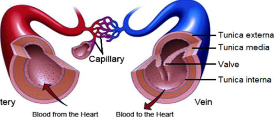
In the picture, the red tube on the left is an artery, which takes blood away from the heart. The blue tube on the right is a vein, which brings blood back to the heart. A mesh of very fine capillaries joins the two. The wall of both an artery and a vein has three layers: an outer layer called the tunica externa, a middle layer of muscle called the tunica media, and an inner layer called the tunica interna. Notice that the muscle layer is thick in the artery and thin in the vein, and that the vein has a valve inside it.
Arteries
Artery: the blood vessel that transports blood from the heart to the various parts of the body.
The heart pushes blood out with great force, so the pressure of blood inside an artery is high. To withstand this pressure, an artery has a thick, strong, highly muscular wall. Blood in an artery is always being driven forward, so arteries have no valves. The largest artery in the human body is the aorta. Think of the main pipe leaving a water tank, which splits into smaller pipes as it reaches each house. In the same way, the aorta divides into many small arteries. These divide further into arterioles, and each arteriole divides again to form a fine network of blood capillaries.
Aorta: the largest artery in the human body. It leaves the left ventricle and divides to give off many small arteries.
Capillaries
Capillaries: the network of fine, thread-like or hair-like blood vessels formed when arterioles divide.
Capillaries are where blood and cells trade materials. Nutrients, oxygen, hormones, enzymes and other substances dissolved in the blood pass from the capillaries to the cells. At the same time, the capillaries receive wastes produced in the cells, such as carbon dioxide and urea, and carry them away so they can reach the excretory organs. A capillary works like a postman who hands letters to every house and collects the letters each house wants to send. After receiving wastes from the cells, capillaries join together to form venules, and venules join to form veins.
Veins
Vein: the blood vessel that transports blood from the various organs of the body to the heart.
The wall of a vein has the same three layers as an artery, but its layer of smooth muscle is thinner, so veins are thin-walled vessels. Veins carry blood rich in carbon dioxide and wastes, collected by the venules, back to the heart. By the time blood reaches a vein it is far from the heart's push, so both its speed and its pressure are low. With low pressure there is a chance that blood could flow the wrong way, for example when it has to climb up from the feet. To stop this, veins have valves at frequent intervals. A valve works like a one-way door. It opens when blood moves towards the heart and shuts when blood tries to slip back.
Valve: a structure found at frequent intervals in veins that lets blood flow in one direction only and prevents it from flowing backwards.
Venules, veins and the venacava all do the same job, which is returning blood to the heart. They differ only in size and number. Venules are the smallest and most numerous, and the venacava is the largest vein of all.
Arteries, Capillaries and Veins Compared
| Feature | Artery | Capillary | Vein |
| Direction of blood | From the heart to the organs | From arterioles to venules | From the organs to the heart |
| Wall | Thick and highly muscular | Fine and hair-like | Thin, with less muscle |
| Blood pressure | High | Falling | Low |
| Valves | Absent | Absent | Present at frequent intervals |
| Main job | Carry blood out towards the cells | Exchange materials with the cells | Return blood to the heart |
What Blood Circulation Is
Now put the three vessels together into one route. Blood leaves the heart through the arteries, reaches the capillaries, and returns to the heart through the veins. This journey never stops. The heart of an adult pumps about 5 to 6 litres of blood per minute, which is roughly a large bucketful travelling round the body every minute.
Blood circulation: the process by which blood is transported from the heart to the various parts of the body and from the various parts of the body back to the heart.
The circulation of blood in the human body can be divided into two kinds:
- Systemic blood circulation, between the heart and the organs of the body
- Pulmonary blood circulation, between the heart and the lungs
Systemic Blood Circulation
Systemic circulation: the circulation in which oxygenated blood from the left ventricle flows through the aorta and its branches (arteries) to the various parts of the body, and deoxygenated blood from these organs returns to the heart through veins.
In systemic circulation, blood travels in this order:
- The left ventricle contracts and sends pure, oxygenated blood out of the heart into the aorta.
- From the aorta the blood passes into smaller arteries and then into arterioles.
- The arterioles lead into capillaries, which supply nutrients, oxygen and other essential substances to every cell.
- The cells use these substances and produce carbon dioxide and other wastes, which the blood carries away through the veins.
- The veins join together to form the venacava.
- The venacava delivers this impure, carbon dioxide-rich blood to the right auricle.

Systemic circulation starts at the left ventricle and ends at the right auricle. It gives the body's cells oxygen and food and brings their wastes back.
Pulmonary Blood Circulation
The blood that reaches the right auricle is low in oxygen and full of carbon dioxide. Before it can be sent round the body again it must be made pure, and that happens in the lungs. The trip between the heart and the lungs is called pulmonary circulation.
Pulmonary circulation: the circulation of blood between the heart and the lungs through the pulmonary artery and pulmonary veins, in which impure blood flows from the right ventricle to the lungs and pure blood flows from the lungs to the left auricle.
In pulmonary circulation, blood travels in this order:
- The right ventricle contracts and the pulmonic valve opens.
- Deoxygenated, carbon dioxide-rich blood flows into the pulmonary artery.
- The pulmonary artery carries it to the network of capillaries surrounding the millions of alveoli in the lungs.
- There the blood leaves its carbon dioxide in the alveoli and picks up oxygen, becoming pure, oxygenated blood.
- The pure blood travels through the pulmonary veins to the left auricle.

Do not assume that an artery always carries pure blood and a vein always carries impure blood. What makes a vessel an artery or a vein is the direction it carries blood. The pulmonary artery carries blood away from the heart, so it is an artery, yet its blood is deoxygenated. The pulmonary veins carry blood towards the heart, so they are veins, yet their blood is oxygenated.
The Complete Journey of Blood
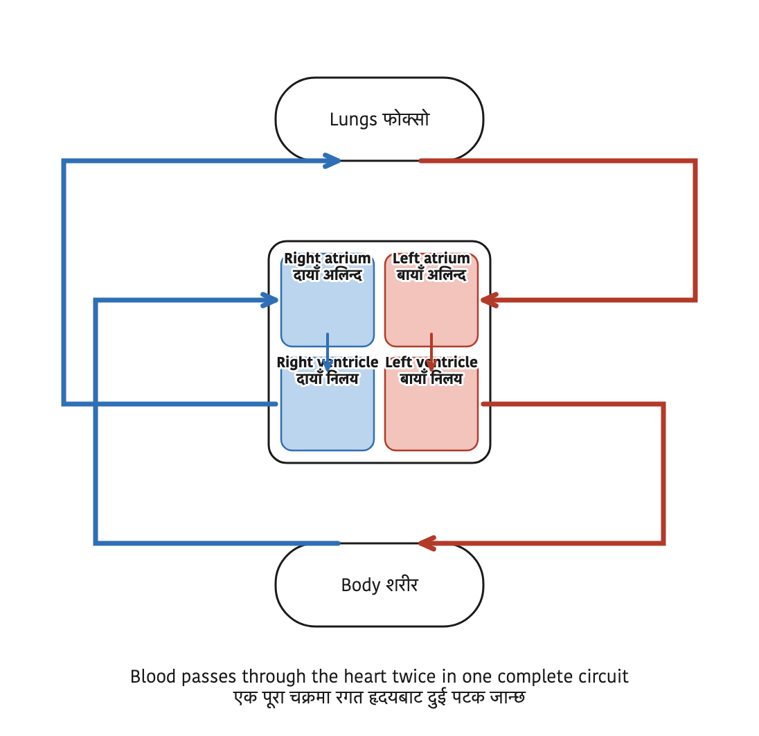
These two circulations are not separate. They are joined at the heart, and each one begins where the other ends. The impure blood that systemic circulation brings to the right auricle passes into the right ventricle and is sent to the lungs by pulmonary circulation. The pure blood that pulmonary circulation brings to the left auricle passes into the left ventricle and is sent round the body by systemic circulation. So the right side of the heart always holds impure blood and the left side always holds pure blood, and blood passes through the heart twice on each complete trip round the body.
| Feature | Systemic circulation | Pulmonary circulation |
| Starts at | Left ventricle | Right ventricle |
| Ends at | Right auricle | Left auricle |
| Vessel leaving the heart | Aorta | Pulmonary artery |
| Vessel returning to the heart | Venacava | Pulmonary veins |
| Blood goes to | The various parts of the body | The lungs |
| Blood leaving the heart | Oxygenated (pure) | Deoxygenated (impure) |
Right ventricle to lungs to left auricle is pulmonary. Left ventricle to body to right auricle is systemic.
The full lesson is waiting for you
The notes are free to read in full. A free account opens the rest of the practice, and tracks your progress and confidence score for every subject. Presentations and videos need approval from Learn Yeti.